. 2015 Aug 1;309(3):R255-76.
doi: 10.1152/ajpregu.00152.2015. Epub 2015 May 27.

Complementary expression and phosphorylation of Cx46 and Cx50 during development and following gene deletion in mouse and in normal and orchitic mink testes

Affiliations

Complementary expression and phosphorylation of Cx46 and Cx50 during development and following gene deletion in mouse and in normal and orchitic mink testes

R-Marc Pelletier et al. Am J Physiol Regul Integr Comp Physiol. .

Erratum in

  • Corrigendum.
    [No authors listed] [No authors listed] Am J Physiol Regul Integr Comp Physiol. 2015 Dec 15;309(12):R1578. doi: 10.1152/ajpregu.zh6-8905-corr.2015. Am J Physiol Regul Integr Comp Physiol. 2015. PMID: 26671929 Free PMC article. No abstract available.

Abstract

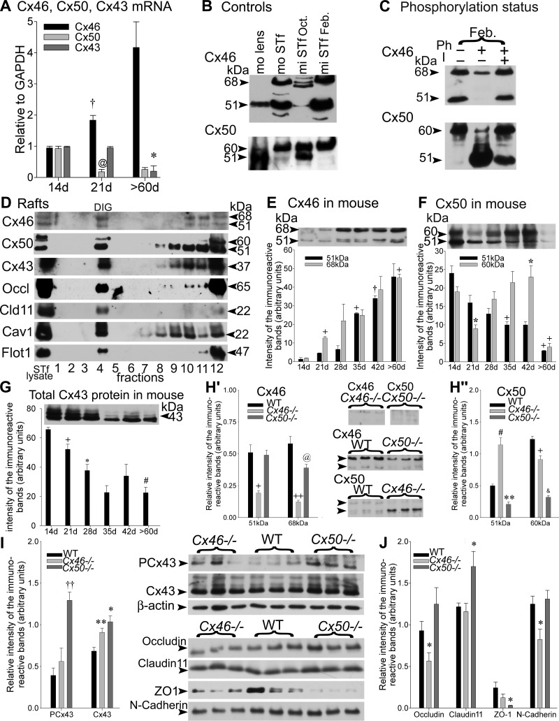
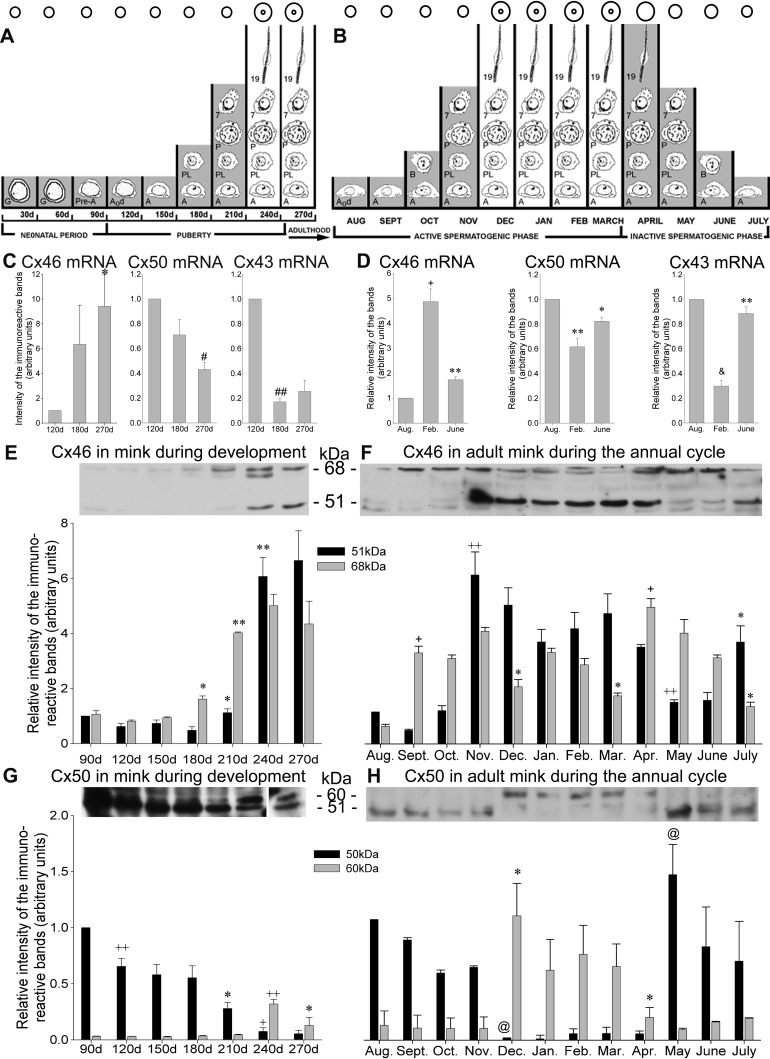
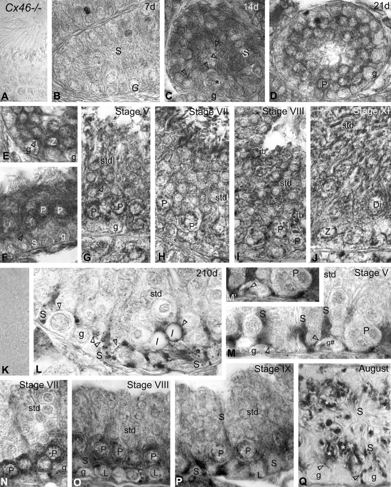
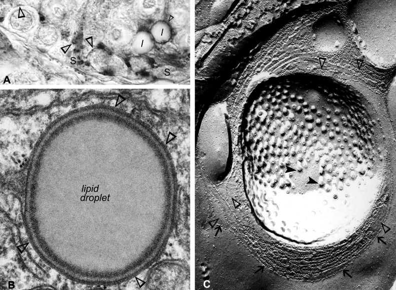
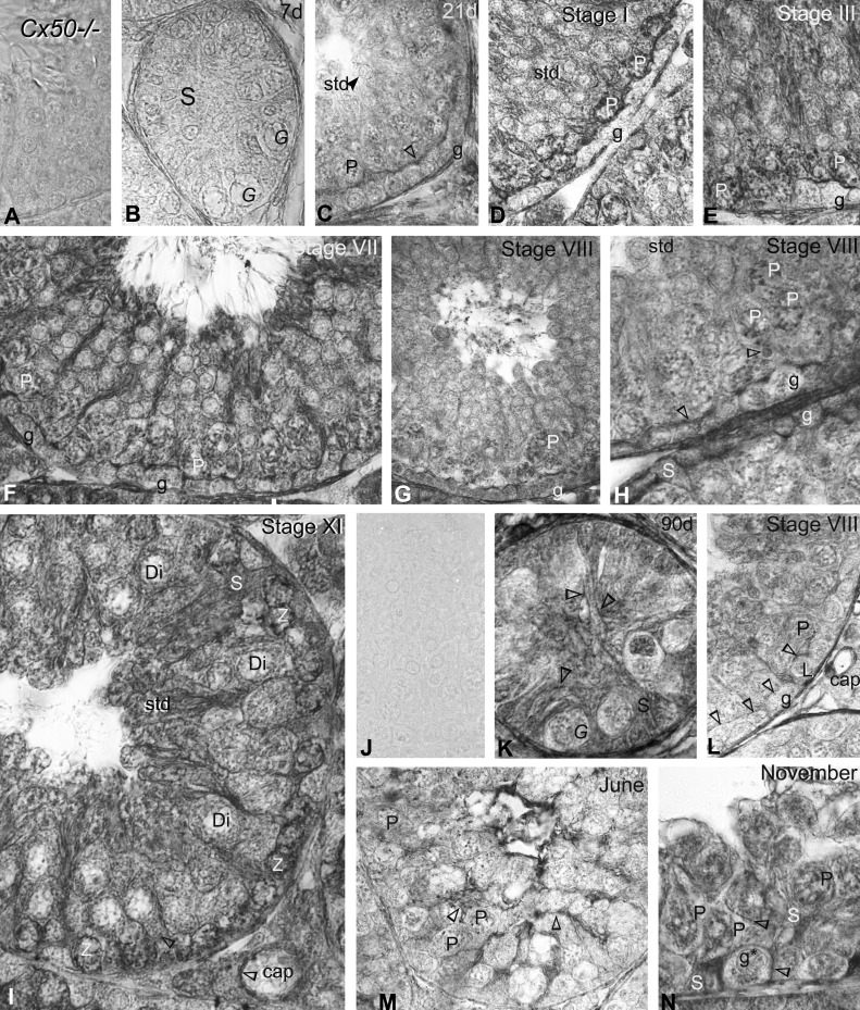
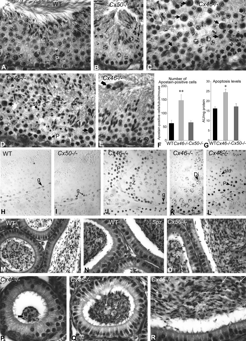
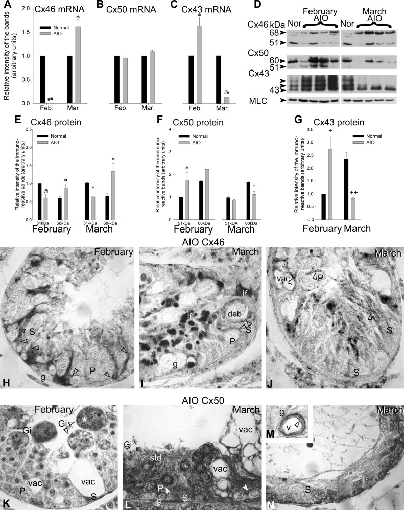
Gap junction-mediated communication helps synchronize interconnected Sertoli cell activities. Besides, coordination of germ cell and Sertoli cell activities depends on gap junction-mediated Sertoli cell-germ cell communication. This report assesses mechanisms underlying the regulation of connexin 46 (Cx46) and Cx50 in mouse testis and those accompanying a "natural" seasonal and a pathological arrest of spermatogenesis, resulting from autoimmune orchitis (AIO) in mink. Furthermore, the impact of deleting Cx46 or Cx50 on the expression, phosphorylation of junction proteins, and spermatogenesis is evaluated. Cx46 mRNA and protein expression increased, whereas Cx50 decreased with adulthood in normal mice and mink. Cx46 mRNA and protein expression increased, whereas Cx50 decreased with adulthood in normal mice and mink. During the mink active spermatogenic phase, Cx50 became phosphorylated and localized to the site of the blood-testis barrier. By contrast, Cx46 was dephosphorylated and associated with annular junctions, suggesting phosphorylation/dephosphorylation of Cx46 and Cx50 involvement in the barrier dynamics. Cx46-positive annular junctions in contact with lipid droplets were found. Cx46 and Cx50 expression and localization were altered in mink with AIO. The deletion of Cx46 or Cx50 impacted on other connexin expression and phosphorylation and differently affected tight and adhering junction protein expression. The level of apoptosis, determined by ELISA, and a number of Apostain-labeled spermatocytes and spermatids/tubules were higher in mice lacking Cx46 (Cx46-/-) than wild-type and Cx50-/- mice, arguing for life-sustaining Cx46 gap junction-mediated exchanges in late-stage germ cells secluded from the blood by the barrier. The data show that expression and phosphorylation of Cx46 and Cx50 are complementary in seminiferous tubules.

Keywords: Sertoli cell; connexin 43; lipid droplet; meiosis; seasonal breeder.


Figures

Fig. 1.
Fig. 1.
A: the samples were subjected to RT-PCR using primers specific for mouse connexin 45 (Cx46), Cx50, and/or Cx43 genes. The Cx46, Cx50, and Cx43 mRNA levels in normal mouse seminiferous tubule-enriched fractions (STfs) during mouse postnatal development are shown. The values are the means ± SE of 3 independent experiments for each age group, expressed in arbitrary units. Cx46 mRNA levels increased significantly from 14 to 21 days (d; †P < 0.03 21 days vs. 14 days) and then raised further by >60 days, in contrast to Cx50 mRNA levels that decreased significantly from 14 to 21 days (@P < 0.02 21 days vs. 14 days) and then remained low by >60 days. The Cx43 mRNA levels decreased steadily throughout development, reaching lowest values by >60 days (*P < 0.05 >60 days vs. 21 days). B: when exposed to the polyclonal Cx46 antibody (Alpha Diagnostic International), a single, intense, ∼51-kDa band was detected in an adult mouse (mo) lens used as a positive control. An additional ∼68-kDa band was, however, recognized in mouse and mink (mi) STfs. The Cx50 antibody detected a 51-kDa-immunoreactive band, accompanied by an ∼60 kDa in the adult mouse lens used as a positive control. The 51- and ∼60-kDa Cx50-immunoreactive bands were detected in mouse STf and mink STf. Oct., October; Feb., February. C: phosphorylation-status studies of Cx46 and Cx50: normal adult mink tubule-enriched fractions in February were incubated in PBS, alone or containing alkaline phosphatase (Ph) in the absence (−) or presence (+) of a phosphatase inhibitor (I). Following treatment, total protein aliquots from each sample were subjected to SDS-PAGE, followed by Western blotting with Cx46 or Cx50 antibodies. Approximately 68- and 51-kDa bands were detected by Cx46 antibodies. Both bands diminished after alkaline phosphatase treatment, revealing the presence of Cx46 phosphorylated forms. The strong 60-kDa band detected by Cx50 antibodies was decreased following alkaline phosphatase treatment, but the 51-kDa band increased, suggesting the presence of phosphorylated forms in the 60-kDa Cx50-immunoreactive band. D: experiments carried out using detergents to isolate the detergent-insoluble glycolipid (DIG) fractions from adult mink tubule-enriched fractions obtained in February: caveolin-1 (Cav1) and flotillin-1 (Flot1) served to identify the DIG fraction by Western blotting. The gap-junction proteins Cx46, Cx50, and Cx43, as well as the tight-junction proteins, occludin (Occl) and claudin 11 (Cld11), were recovered in the DIG fraction. E–G: representative Western blot analyses and quantification of individual levels of the 51- and ∼68-kDa (E) Cx46-immunoreactive bands and (F) Cx50-immunoreactive bands and (G) total Cx43, measured in mouse tubule-enriched fractions during development, are shown. The values are the means ± SE of 3 independent experiments expressed in arbitrary units. E: the changes in Cx46 protein levels were significant as follows: 51 kDa, +P < 0.01 35 days vs. 28 days and †P < 0.03 42 days vs. 35 days; 68 kDa, +P < 0.01 21 days vs. 14 days and >60 days vs. 21 days. F: the following changes in Cx50 levels were significant: 51 kDa, +P < 0.01 35 days vs. 14 days and >60 days vs. 42 days; 60 kDa, *P < 0.05 21 days vs. 14 days and 42 days vs. 21 days and +P < 0.01 60 days vs. 42 days. G: the following changes in Cx43 protein levels were significant: +P < 0.01 21 days vs. 14 days, *P < 0.05 28 days vs. 21 days, and #P < 0.0001 >60 days vs. 14 days. H–J: compensatory studies carried out in mice lacking Cx46 (Cx46−/−), wild-type (WT) mice, and Cx50−/− tubule-enriched fractions. The values shown are the means ± SE of 3 independent experiments and are expressed in arbitrary units. H′ and H“: representative Western blots, accompanied by histograms of the quantification of Cx46 and Cx50 levels in each animal group, are presented. As expected, the intensity of the Cx46 (H′)- and Cx50 (H”)-immunoreactive band levels was reduced to insignificant traces in Cx46−/− (51 kDa band: +P < 0.01 Cx46−/− vs. WT; 68 kDa band: ++P < 0.001 Cx46−/− vs. WT) and Cx50−/− (51 kDa band: **P < 0.005 Cx50−/− vs. WT; 60 kDa band: &P < 0.0005 Cx50−/− vs. WT) mice, respectively. H′: the 68-kDa Cx46 band levels decreased significantly (@P < 0.02 Cx50−/− vs. WT) in Cx50−/− mice. H“: the 51-kDa Cx50-immunoreactive band levels increased significantly (#P < 0.0001 Cx46−/− vs. WT), whereas 60 kDa levels were reduced (+P < 0.001 Cx46−/− vs. WT) in the Cx46−/− mice. Representative Western blots of (I) PCx43 (phosphorylated in serine 368), Cx43, and (J) occludin, claudin 11, zona occulden protein 1 (ZO-1), and N-cadherin levels, accompanied by histograms of the quantification of the levels measured for each junction protein in WT, Cx46−/−, and Cx50−/− mice tubule-enriched fractions. The following changes in the levels of each junction protein were significant: ††P < 0.003 PCx43 Cx50−/− vs. WT; **P < 0.005 Cx43 Cx46−/− vs. WT; *P < 0.05 Cx43 Cx50−/− vs. WT; *P < 0.05 occludin Cx46−/− vs. WT; *P < 0.05 claudin 11 Cx50−/− vs. WT; *P < 0.05 ZO-1 Cx50−/− vs. WT; *P < 0.05 N-cadherin Cx46−/− vs. WT.
Fig. 2.
Fig. 2.
Variations in the Cx46, Cx50, and Cx43 mRNA and protein levels in mink STfs during postnatal development and the annual reproductive cycle. A calendar of the germ cell population recorded during the (A) postnatal development and (B) annual seasonal reproductive cycle in mink seminiferous tubule cross-sections is provided. The mink breed in the 2nd and 3rd wk of March. The pups are born in April–May. Each month is represented by a vertical column in the 2 diagrams. The columns either span (A) from birth to adulthood by 270 days after birth or (B) cover the 12-mo seasonal reproductive cycle of the adult mink. The shading refers to time periods when the blood-testis barrier is permeable to vascularly infused permeability tracers. Conversely, the months when the barrier is competent in blocking tracers are not shaded. The inner circles apposed atop indicate the presence of a lumen. G, gonocytes; Pre-A, pre-type A spermatogonia; A0d, type A0 spermatogonia dividing; A, type A spermatogonia; PL, preleptotene spermatocyte; P, pachytene spermatocyte; 7, step 7 spermatid; 19, step 19 spermatid; B, type B spermatogonia. After a 90-day neonatal period, puberty encompasses the colonization of the tubules by type A spermatogonia until reaching the 1st spermatozoa in the epididymis, ∼250 days after birth. This is followed, first by an ”active“ and then by an ”inactive“ phase of the annual reproductive cycle in the adult mink. A reduction in mitotic and meiotic activities yields spermatids in reduced abundance. By August (AUG), the division of spermatogonial stem cells (A0d) marks the onset of the active phase of the annual seasonal reproductive cycle [after Pelletier (68) and modified by Pelletier et al. (82)]. The individual Cx46, Cx50, and Cx43 mRNA levels, measured by RT-PCR in tubule-enriched fractions, are plotted in arbitrary units during (C) development and (D) the annual seasonal reproductive cycle in the adult mink. The values are the means ± SE of 3 independent experiments and are normalized to the 120-day values (C) and August values (D). C: the Cx46 mRNA levels increased significantly (*P < 0.05 270 days vs. 120 days); however, Cx50 decreased (#P < 0.0001 270 days vs. 120 days) by adulthood. Similarly, Cx43 fell significantly (##P < 0.00001 180 days vs. 120 days) by 180 days after birth and then remained low in the adult. D: during the seasonal reproductive cycle, Cx46 mRNA levels were highest in February (+P < 0.001 Feb. vs. Aug.) but then decreased significantly by June (**P < 0.005 June vs. Feb.). By contrast, Cx50 mRNA levels were lowest in February (**P < 0.005 Feb. vs. Aug.) before increasing significantly by June (*P < 0.05 June vs. Feb.). The Cx43 levels were lowered significantly by February (&P < 0.0005 Feb. vs. Aug.) but increased significantly by June (**P < 0.005 June vs. Feb.). E and F: representative Western blots with histograms showing the monthly changes in Cx46 protein levels in tubule-enriched fractions during (E) development and (F) the annual reproductive cycle in mink. Quantification of the levels of the 51- and 68-kDa Cx46-immunoreactive bands is provided. The values are the means ± SE of 3 independent experiments expressed in arbitrary units and normalized to the 51-kDa band in 90-day-old mink for studies on development and normalized to August during the annual reproductive cycle. The changes in Cx46 protein levels were significant during (E) development (51 kDa band: *P < 0.05 210 days vs. 180 days and **P < 0.005 240 days vs. 210 days; 68 kDa band: *P < 0.05 180 days vs. 150 days and **P < 0.005 210 days vs. 180 days) and (F) the seasonal reproductive cycle (51 kDa band: ++P < 0.001 Nov. vs. Sept. and May vs. April and *P < 0.05 July vs. June; 68 kDa: +P < 0.01 Sept. vs. Aug. and April vs. March and *P < 0.05 Dec. vs. Nov., March vs. Feb., and July vs. June). G and H: representative Western blots of Cx50 in mink tubule-enriched fractions during (G) development and (H) the annual reproductive cycle. The quantification of the 51- and 60-kDa Cx50-immunoreactive band levels are plotted monthly. The values are the means ± SE of 3 independent experiments expressed in arbitrary units and normalized to the 51-kDa band in the 90 day old during development and to August during the annual reproductive cycle. The following changes in Cx50 levels were significant during development [G; 51 kDa band (black bars): ++P < 0.001 120 days vs. 90 days, *P < 0.05 210 days vs. 180 days, and +P < 0.01 240 days vs. 210 days; 60 kDa band (gray bars): ++P < 0.001 240 days vs. 210 days and *P < 0.05 270 days vs. 240 days] and the annual reproductive cycle (H; 51 kDa band: @P < 0.02 Dec. vs. Nov. and May vs. April; 60 kDa band: *P < 0.05 Dec. vs. Nov. and April vs. March).
Fig. 3.
Fig. 3.
Immunoperoxidase labeling with anti-Cx46 in (A–J) mice and (K–Q) mink. A: no reaction product is detected with Cx46 antibodies used on Cx46−/− mice testis sections. B: Cx46 labeling was virtually undetectable in this 7-day-old testis section. G, undifferentiated spermatogonia; S, Sertoli cell. C: Sertoli cell plasma membranes, whether facing each other near the center of this developing tubule or engaged in Sertoli cell-to-Sertoli cell and Sertoli cell-to-germ cell (*) contacts near the basal 3rd, are heavily labeled (open arrowheads) by 14 days. g, spermatogonia. D–F: the colonization by spermatocytes of the seminiferous tubules and canalization of a lumen are seen in this 21-day-old mouse testis section. The open arrowheads point to labeling at (F) Sertoli cell plasma membranes and (E) intercellular contacts. The endoplasmic reticulum and Golgi apparatus of (E) zygotene (Z) and (D and F) pachytene (P) spermatocytes are heavily labeled. G–J: as well, Sertoli plasma membranes (open arrowhead in G) and the endoplasmic reticulum and Golgi apparatus of pachytene, zygotene, and diplotene (Di) spermatocytes are labeled in adult mice. The stages of the cycle of seminiferous epithelium appear at the top of the micrographs. std, spermatid; rb, residual body. K: no immunostaining is detected in Cx46 controls done on a normal adult mink testis section obtained in February when using the primary or secondary antibody alone. L: Cx46 immunolabeling is shown in a 210-day-old and (E–I) adult mink testes in (M–P) February and (Q) August. Labeling (open arrowheads) is identified in variously sized vacuoles scattered within the trunk of Sertoli cells, as well as in Sertoli cell contacts with the germ cells (spermatogonia; pachytene spermatocytes). Lipid droplets (l), near the base of Sertoli cells, are surrounded by an intense, Cx46-positive halo (open arrowheads). M–P: in February, Cx46 labeling is seen (open arrowheads) in regions of Sertoli cells and in their contacts between themselves and with spermatogonia, leptotene (L), and pachytene spermatocytes during the different stages of the mink seminiferous cycle, appearing in roman numerals atop the figures. M′: a higher magnification of Cx46 labeling in the Golgi region of the spermatogonium, identified g# in M. The endoplasmic reticulum and the Golgi zone of the leptotene and pachytene spermatocytes are labeled. Cx46 labeling decreased in pachytene spermatocytes from stage VIII (O) to stage IX (P). Q: during the seasonal testicular regression, Cx46 labels (arrowheads) thin Sertoli cell processes, some of which are in contact with spermatogonia and pachytene spermatocytes, remaining in seminiferous tubules by August. Lipofuscin pigments are identified (asterisks) in regressed Sertoli cells. Original magnification, ×860 (A–M and N–Q); ×980 (M′).
Fig. 4.
Fig. 4.
A: this is a higher magnification of a portion of the field shown in Fig. 3L, in which the attention is drawn here to Cx46 labeling, detected (open arrowheads) not only in Sertoli cell-to-germ cell junctions but also in annular junctions found either wrapped around a lipid droplet or scattered within the trunk of the Sertoli cell. B: electron microscopy of a thin section of an annular junction, corresponding to the one shown in A, is shown, establishing contacts with the surface of a lipid droplet. C: a corresponding image in electron microscopy of freeze fracture. In this micrograph, an impressive circular array of Sertoli cell annular gap junctions (open arrowheads), intercalated amongst strands of tight-junctional particles (arrows), is seen surrounding a cluster of filipin-cholesterol complexes (closed arrowheads). Original magnification, ×1,000 (A); ×1,000,000 (B); ×116,000 (C).
Fig. 5.
Fig. 5.
Immunoperoxidase labeling with anti-Cx50 in (A–I) mice and (J–N) mink testis sections. A: no reaction product was detected when Cx50 antibodies were used on Cx50−/− mice testis sections. B: no reaction product was detectable in 7-day-old mice testis sections. C: this 21-day-old mouse testis section shows Cx50 labeling (open arrowhead) in the cell contacts settled at the site of the blood-testis barrier. D–H: at this site, Cx50 labeling is seen in cell contacts established above spermatogonia and young spermatocytes in the basal 3rd of the seminiferous epithelium during the stage cycles in adult mice. D–J: in addition, zygotene and pachytene spermatocytes show intense Cx50 labeling. I: the wall of the capillary (cap) is labeled. J: Cx50 control, done using either the 1st or the 2nd antibody in adult mink testis sections obtained in February, shows no immunostaining. K: the Sertoli cell membranes show delicate labeling (open arrowheads) in a 60-day-old mink. L: in February, the distribution of Cx50 (open arrowheads) above spermatogonia and young spermatocytes in the tubules closely coincides with that of junctional complexes established at the site of the blood-testis barrier in mink. M: the lumen of the seminiferous tubule is collapsed in June during testicular regression, and contacting Sertoli cell plasma membranes are labeled; however, the distribution of Cx50 labeling no longer coincides with that of the junctional complexes at the site of the blood-testis barrier. N: contacts between Sertoli cells and spermatogonia pachytene spermatocytes are labeled in November. As well, the Golgi zone of the spermatogonium identified g*; spermatid contains minuscule Cx50-positive dots. Original magnification, ×860.
Fig. 6.
Fig. 6.
A–E: Bouin's-fixed periodic acid Schiff (PAS)-stained paraffin testis sections from (A) normal, >60-day-old WT, (B) Cx50−/−, and (C–E) Cx46−/− adult mice. A and B: spermatogenesis appeared normal in WT and Cx50−/− mice testis sections, in which pachytene spermatocytes and dividing secondary spermatocytes are identified (II*). C and D: however, Cx46−/− testis sections showed numerous tubules with plentiful apoptotic cells, particularly pachytene and diplotene spermatocytes, involved in the meiotic division (arrows). In other stages of the cycle, apoptotic spermatids were observed. E: the lumen was collapsed in some tubules, whereas in others, cellular debris or clusters of apoptotic cells (wide arrow) were observed. F: a histogram of the quantification of immunoperoxidase Apostain-labeled cells is presented. The bars represent the means ± SE of Apostain-positive cells, counted in 25 tubules from 3 different mice per experimental group. Apostain-labeled cells were significantly more numerous in Cx46−/− than in WT seminiferous tubules (**P < 0.005 Cx46−/− vs. WT). The number of labeled cells in WT and Cx50−/− mice did not differ significantly. G: a histogram of nucleosome release measured by cell death detection ELISA in the cytoplasmic fraction of WT, Cx46−/−, and Cx50−/− mice tubule-enriched fractions obtained is shown. The data are expressed in optical density at 410 nm, and the bars represent the means ± SE of measurements in 3 different mice per experimental group. The increase in nucleosome release in Cx46−/− is significantly different (*P < 0.05) compared with WT but not between WT and Cx50−/−. H–L: immunoperoxidase labeling of cells in apoptosis with antibody F7-26 (Apostain) is shown. Apostain-positive spermatogonia, zygotene, pachytene, and diplotene spermatocytes and spermatids were found in comparable quantities in (H) WT and (I) Cx50−/− mice. J–L: in Cx46−/− mice, Apostain labeling involved spermatogonia and spermatids. In addition, zygotene, pachytene, and diplotene spermatocytes were labeled in larger quantities in Cx46−/− than in WT and Cx50−/− mice. M–R: Bouin's-fixed PAS-stained epididymis paraffin sections from (M and N) WT, (O) Cx50−/−, and (P–R) Cx46−/− mice are shown. The (M) body and (N and O) tail of the epididymides of (M and N) WT and (O) Cx50−/− exhibited similar histological features, and both contained spermatozoa (Spz) in comparable amounts. However, (P and Q) the epididymides from Cx46−/− showed spermatozoa in reduced quantities. P: a cluster of apoptotic cells, reminiscent of the one shown in the testis in Fig. 3E, is identified (wide arrow). Apoptotic spermatocytes and other young germ cells are identified by arrows in the (P) head, (Q) body, and (R) tail of Cx46−/− mice epididymides. Original magnification, ×720 (A–E); ×860 (H and I and K–O); ×950 (J and P–R).
Fig. 7.
Fig. 7.
A–C: the seminiferous tubule-enriched samples were subjected to RT-PCR. The (A) Cx46, (B) Cx50, and (C) Cx43 mRNA levels measured in normal adult mink were compared with those in mink with autoimmune orchitis (AIO) in February and March (Mar.). The data in arbitrary units are expressed as the means ± SE of 3 independent experiments and normalized to the normal value for each month. The differences measured between normal mink and mink with AIO were statistically significant for (A) Cx46 mRNA (##P < 0.00001 Feb. AIO vs. Feb. Normal and *P < 0.05 Mar. AIO vs. Mar. Normal) and for (C) Cx43 mRNA (*P < 0.05 Feb. AIO vs. Feb. Normal and ##P < 0.00001 Mar. AIO vs. Mar. Normal). B: the differences in Cx50 mRNA levels measured in normal mink and mink with AIO show no significant difference. Representative Western blots of (D) Cx46, Cx50, and Cx43 in normal (Nor) adult mink and mink with AIO in February and March are shown. The quantification of (E) Cx46, (F) Cx50, and (G) Cx43 protein levels, measured in normal mink and mink with AIO, is provided. The values are the means ± SE of 4 independent experiments, expressed in arbitrary units, normalized to the 51-kDa Cx46 for February, the 51-kDa Cx50 for February, and total Cx43 for February. The myosin light chain (MLC) was used as the internal loading control and did not change significantly in the different experimental conditions of the study. Significant differences were measured in the junction protein levels between normal mink and mink with AIO: Cx46, February, 51 kDa band: @P < 0.02 AIO vs. Normal, 68 kDa band: *P < 0.05 AIO vs. Normal; March, 51 kDa band: *P < 0.05 AIO vs. Normal, 68 kDa band: *P < 0.05 AIO vs. Normal; Cx50, February, 51 kDa band: *P < 0.05 AIO vs. Normal; March, 60 kDa band: †P < 0.03 AIO vs. Normal; Cx43: February, +P < 0.01 AIO vs. Normal; March, ++P < 0.001 AIO vs. Normal. Immunoperoxidase labeling with anti-Cx46 in (H) February and (I and J) March and with anti-Cx50 in (K) February and (L–N) March of orchitic adult mink testis paraffin sections, respectively. H: Cx46 labeling (open arrowheads) is seen amongst Sertoli cells and Sertoli cell–Sertoli cell, Sertoli cell–spermatogonia, and pachytene spermatocytes contacts. I and J: the membrane of vacuoles (vac) remaining behind the exfoliation of germ cells is Cx46 positive. I: some of these vacuoles contain cellular debris (deb). IF, lipofuscin pigment. K: giant cells remaining within or being released from the seminiferous epithelium are identified (Gi). The cells within the giant cell (Gi*) show signs of apoptosis. L: Cx50 labeling (open arrowhead) is observed amongst Sertoli cells, Sertoli cell contacts to germ cells, as well in the perinuclear zone of pachytene spermatocytes in the early phase of AIO but not in (N) tubules, where destruction is massive in the late phase of the disease. M: Cx50 labeling (open arrowhead) is seen in the wall of a blood vessel (v). Original magnification, ×860.

Similar articles

Cited by

References

    1. Abraham V, Chou ML, George P, Pooler P, Zaman A, Savani R, Koval M. Heterocellular gap junctional communication between alveolar epithelial cells. Am J Physiol Lung Cell Mol Physiol 280: L1085–L1093, 2001. - PubMed
    1. Akpovi CD, Pelletier RM. A revised and improved method for the isolation of seminiferous tubule-enriched fractions that preserves the phosphorylated and glycosylated forms of proteins. In: Human Embryogenesis Methods and Protocols, edited by Lafond J, Vaillancourt C. Totowa, NJ: Humana, Springer Protocols, 2009, p. 159–168. - PubMed
    1. Akpovi CD, Yoon SR, Vitale ML, Pelletier RM. The predominance of one of the SR-BI isoforms is associated with increased esterified cholesterol levels not apoptosis in mink testis. J Lipid Res 47: 2233–2247, 2006. - PubMed
    1. Alcala J, Katar M, Maisel H. Lipid composition of chick lens fiber cell gap junctions. Curr Eye Res 2: 569–578, 1982. - PubMed
    1. Baldo GJ, Gong X, Martinez-Wittimghan FJ, Kumar NM, Gilula NB, Mathias RT. Gap junctional coupling in lenses from alpha(8) connexin knockout mice. J Gen Physiol 118: 447–456, 2001. - PMC - PubMed

Publication types

LinkOut - more resources