. 2015 Aug;138(Pt 8):2219-33.
doi: 10.1093/brain/awv142. Epub 2015 May 27.

Dissecting the phenotypes of Dravet syndrome by gene deletion

Affiliations

Dissecting the phenotypes of Dravet syndrome by gene deletion

Moran Rubinstein et al. Brain. 2015 Aug.

Abstract

Neurological and psychiatric syndromes often have multiple disease traits, yet it is unknown how such multi-faceted deficits arise from single mutations. Haploinsufficiency of the voltage-gated sodium channel Nav1.1 causes Dravet syndrome, an intractable childhood-onset epilepsy with hyperactivity, cognitive deficit, autistic-like behaviours, and premature death. Deletion of Nav1.1 channels selectively impairs excitability of GABAergic interneurons. We studied mice having selective deletion of Nav1.1 in parvalbumin- or somatostatin-expressing interneurons. In brain slices, these deletions cause increased threshold for action potential generation, impaired action potential firing in trains, and reduced amplification of postsynaptic potentials in those interneurons. Selective deletion of Nav1.1 in parvalbumin- or somatostatin-expressing interneurons increases susceptibility to thermally-induced seizures, which are strikingly prolonged when Nav1.1 is deleted in both interneuron types. Mice with global haploinsufficiency of Nav1.1 display autistic-like behaviours, hyperactivity and cognitive impairment. Haploinsufficiency of Nav1.1 in parvalbumin-expressing interneurons causes autistic-like behaviours, but not hyperactivity, whereas haploinsufficiency in somatostatin-expressing interneurons causes hyperactivity without autistic-like behaviours. Heterozygous deletion in both interneuron types is required to impair long-term spatial memory in context-dependent fear conditioning, without affecting short-term spatial learning or memory. Thus, the multi-faceted phenotypes of Dravet syndrome can be genetically dissected, revealing synergy in causing epilepsy, premature death and deficits in long-term spatial memory, but interneuron-specific effects on hyperactivity and autistic-like behaviours. These results show that multiple disease traits can arise from similar functional deficits in specific interneuron types.

Keywords: Dravet syndrome; Nav1.1; interneurons; parvalbumin; sodium channel; somatostatin.


Figures

None
Dravet Syndrome is a complex neuropsychiatric disease caused by mutations in SCN1A, which encodes the Nav1.1 channel. Rubinstein et al. delete Scn1a in parvalbumin- or somatostatin-expressing interneurons in mice, and show that the former manipulation induces epilepsy and autistic-like behaviours, the latter epilepsy and hyperactivity, while both together impair cognition.
Figure 1
Figure 1
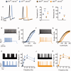
Deletion of Nav1.1 results in reduced excitability in layer V cortical interneurons. (A) Examples of action potentials recorded in PV- and SST-expressing layer V cortical interneurons. Scn1a+/+ are depicted in black. (B and C) Threshold (B) and rheobase for action potentials (C) in cortical PV (blue) and SST (yellow) neurons. Action potentials were evoked by 10 ms depolarizing current injection. (D–G) Firing of cortical neurons with selective deletion of Nav1.1 in response to 1-s long depolarizing current injection at the indicated intensities. (D) Sample traces of whole-cell current-clamp recordings of PV-expressing neurons in response to injection of 240 pA current. (E) Average number of action potentials in response to 1 s depolarizing current injection at the indicated intensity (n = 15–19). (F) Sample traces of whole-cell current-clamp recordings of SST-expressing neurons in response to injection of 160 pA current (n = 15–17). (G) Average number of action potentials in response to 1 s depolarizing current injection at the indicated intensity. (HK) Failure rates for action potential generation in cortical neurons with selective deletion of Nav1.1. Per cent failure of action potentials in PV (H and I) and SST (J and K) interneurons. A train of 100 pulses, each 10 ms long at the minimal current required to trigger action potentials, was given at the indicated frequencies. Sample traces for 100 pulses at 10 Hz are shown (H and J) n = 15–17. Statistical analysis was done using Student’s t-test. *P < 0.05, **P < 0.01, ***P < 0.001.
Figure 2
Figure 2
Deletion of Nav1.1 in PV neurons, but not in SST neurons, alters the excitation/inhibition balance. Spontaneous IPSC and spontaneous EPSC were recorded in layer V pyramidal neurons from mice with specific deletion of Nav1.1 in PV or SST mice. (AD) PV-DS mice. Example traces of spontaneous IPSC (A) and cumulative histogram of inter-event intervals (B, P = 0.05, Kolmogorov-Smirnov test) with mean values ± SEM (inset) of spontaneous IPSC frequency. Spontaneous IPSC amplitude was 35.9 ± 3.1 pA in PVCre Scn1a+/+ and 36 ± 3.4 pA in PVCre Scn1afl/+, P = 0.49. n = 21–24. Example traces of spontaneous EPSC (C) and cumulative histogram of interevent intervals (D, P = 0.038, Kolmogorov-Smirnov test) with means ± SEM (inset) of sEPSC frequency. Spontaneous EPSC amplitude was 25.3 ± 1 pA in PVCre Scn1a+/+ and 26.7 ± 2.2 pA in PVCre Scn1afl/+, P = 0.28, n = 19–20. (EH) SST-DS mice. Example traces of spontaneous IPSC (E) and cumulative histogram of interevent intervals (F) with means ± SEM (inset) of spontaneous IPSC frequency. Spontaneous IPSC amplitude was 39.5 ± 5.3 pA in SSTCre Scn1a+/+ and 39.4 ± 3 pA in SSTCre Scn1afl/+, P = 0.48. n = 25. Example traces of spontaneous EPSC (G) and cumulative histogram of interevent intervals (H) with means ± SEM (inset) of spontaneous EPSC frequency. Spontaneous EPSC amplitude was 28 ± 1.4 pA in SSTCre Scn1a+/+ and 27.9 ± 1.3 pA in and SSTCre Scn1afl/+, P = 0.37, n = 19–21. Statistical analysis was done using Student’s t-test. *P < 0.05, **P < 0.01.
Figure 3
Figure 3
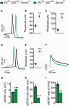
Deletion of Nav1.1 results in reduced excitability of CA1 stratum oriens hippocampal interneurons. (A–C) Action potentials evoked by direct depolarization of cell body via the patch pipette. (A) Examples of action potentials. (B and C) Threshold (B) and rheobase (C) of action potentials, n = 7. (D–G) Properties of synaptically evoked action potentials and near-threshold EPSPs in stratum oriens neurons. Stimulating electrode was placed in the alveus and the stimulation strength was set to produce firing probability of 50% during trains of 10 stimuli at 1 Hz. Representative action potentials (D) and mean values ± SEM for threshold for action potential generation (E). (F–I) Parameters for near-threshold EPSPs. Examples of EPSPs (F). Mean values ± SEM for EPSP amplitude (G), duration (H) and time integral (I), n = 7–9. To control for changes in properties of voltage-dependent currents activated by EPSPs of different amplitude, we examined the duration of EPSPs with similar amplitude. These were also of shorter duration in PV&SST-DS (Supplementary Fig. 2E and F), indicating that loss of Nav1.1 currents, and not voltage-dependent changes in the kinetics of the EPSP due to its higher amplitude, cause reduced amplification. Statistical analysis was done using Student’s t-test. **P < 0.01, ***P < 0.001.
Figure 4
Figure 4
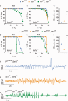
Epileptic phenotype of mice with deletion of Nav1.1 in PV- and SST-expressing interneurons. (A–C) Epileptic phenotypes of mice with heterozygous expression of Cre. (AB) Percentage of mice remaining free of behavioural seizures (SZ) at the indicated body core temperatures at postnatal Day 21 (P21, A) and postnatal Day 35 (P35, B). Selective deletion of Nav1.1 in PV-expressing neuron (blue, n = 10), SST-expressing neurons (yellow, n = 8), or combined deletion of Nav1.1 in both PV and SST expressing neurons (green, n = 17). (C) Mean values ± SEM for seizure duration at postnatal Day 35. (DF) Epileptic phenotypes of mice with homozygous expression of PV Cre. Mean values ± SEM for the percentage of mice remaining free of behavioural seizures (SZ) at the indicated body core temperatures at postnatal Day 21 (D) and postnatal Day 35 (E). (F) Mean values ± SEM for seizure duration at postnatal Day 35. Statistical analysis was done using one-way ANOVA, **P < 0.01. PVCre/Cre-DS, n = 9, SSTCre/Cre-DS, n = 5, PVCre/Cre&SST-DS, n = 8. The epileptic phenotype SSTCre, Scn1afl/+was indistinguishable from that of SSTCre/Cre, Scn1afl/+ (P > 0.05). Similarly the phenotype of PVCre/Cre, SSTCre, Scn1afl/+ was indistinguishable from that of PVCre/Cre, SSTCre/Cre, Scn1afl/+ (n = 4 for each) and these data were pooled. Scn1a+/+ mice of either genotype do not have seizures at the tested temperatures (n = 3–7 for each genotype). (G–I) Examples of EEG recordings from cortical lead depicting a spontaneous electrographic seizure in PVCre/Cre-DS (G), SSTCre-DS (H) and PVCre/Cre&SST-DS (I).
Figure 5
Figure 5
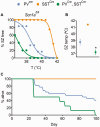
Epileptic phenotype of mice with selective deletion of both alleles encoding Nav1.1. (AC) Epileptic phenotypes of Scn1afl/fl mice. (A) Percentage of mice remaining free of behavioural seizures (SZ) at the indicated body core temperatures at postnatal Day 21. (B) Means values ± SEM for the temperature of seizure induction. (C) Survival plots for PVCre, Scn1afl/fl mice (n = 22, blue), SSTCre, Scn1afl/fl mice (n = 15, yellow) and PVCre, SSTCre, Scn1afl/fl mice (n = 8, green). Statistical analysis was done using one-way ANOVA. *P < 0.05.
Figure 6
Figure 6
Selective deletion of Nav1.1 in SST-expressing neurons causes hyperactivity. Distance moved in the open field test for PV (A, n = 8), SST (B, n = 6), and PV&SST mice (C, n = 10). The behaviour of SSTCre and SSTCre/Cre mice was indistinguishable in all the tests performed and the data were pooled together. Statistical analysis was done using Student’s t-test. *P < 0.05.
Figure 7
Figure 7
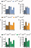
Selective deletion of Nav1.1 in PV-expressing neurons causes social deficit. Three-chamber test of social interaction for PV (A and B, n = 8–9), SST (C and D, n = 11–14) and PV&SST (E and F, n = 6–7) mice. Time in each chamber (A, C and E) and total interaction time (B, D and F): O = object (an empty small mouse cage); C = centre; M = mouse (a stranger mouse in an identical small cage). Statistical analysis was done using Student’s t-test., *P < 0.05, **P < 0.01, ***P < 0.001.
Figure 8
Figure 8
Cognitive phenotype of mice with selective deletion of Nav1.1. Context-dependent fear conditioning test in PVCre (A, n = 16-19 for Scn1a+/+ and Scn1afl/+, respectively), SST (B, n = 10), PV&SST mice (C, n = 10) and PVCre/Cre&SST mice (D, n = 7–9). Freezing behaviour of PVCre (n = 6–9) were indistinguishable from that of PVCre/Cre (n = 10) and these data were pooled in A. Basal = freezing time (%) during exploration of a cage containing an electrical grid as a floor and markings for identification; training = freezing time (%) during presentation of a 0.6 mA, 2 s long foot shock; 30 min = freezing time (%) upon return to the same cage after 30 min but without shock; 24 h = freezing time (%) upon return to the same cage after 24 h but without shock. (E) In the Barnes circular maze, both PVCre/Cre&SST-DS mice and littermate controls decreased their latency to the target hole similarly over the 4-day repeated training trials. (F) Latency to the target hole during the probe trial on the fifth day (n = 6 for each genotype). (G) In the novel object recognition test PVCre/Cre&SST-DS mice and littermate controls had normal recognition memory for a preconditioned object (F = familiar), which was presented 2 min before the test, and spent more time with the novel object (N = novel). (H) The normalized ratio of time spent with the familiar object divided by time spent with the novel object (n = 10–11 for each genotype). (I) Y-maze task: spontaneous alternation behaviour during a 10-min session (n = 5 for each genotype). Statistical analysis was done using Student’s t-test. *P < 0.05, **P < 0.01.

Comment in

  • Phenotypes of Dravet Syndrome.
    Garcia-Sosa R, Laux LC. Garcia-Sosa R, et al. Pediatr Neurol Briefs. 2016 May;30(5):28. doi: 10.15844/pedneurbriefs-30-5-1. Pediatr Neurol Briefs. 2016. PMID: 27617639 Free PMC article.

References

    1. Belforte JE, Zsiros V, Sklar ER, Jiang Z, Yu G, Li Y, et al. Postnatal NMDA receptor ablation in corticolimbic interneurons confers schizophrenia-like phenotypes. Nat Neurosci 2010; 13: 76–83. - PMC - PubMed
    1. Carter BC, Giessel AJ, Sabatini BL, Bean BP. Transient sodium current at subthreshold voltages: activation by EPSP waveforms. Neuron 2012; 75: 1081–93. - PMC - PubMed
    1. Cheah CS, Yu FH, Westenbroek RE, Kalume FK, Oakley JC, Potter GB, et al. Specific deletion of NaV1.1 sodium channels in inhibitory interneurons causes seizures and premature death in a mouse model of Dravet syndrome. Proc Natl Acad Sci USA 2012; 109: 14646–51. - PMC - PubMed
    1. Coulter DA, Yue C, Ang CW, Weissinger F, Goldberg E, Hsu F-C, et al. Hippocampal microcircuit dynamics probed using optical imaging approaches. J Physiol 2011; 589: 1893–903. - PMC - PubMed
    1. Do-Monte FH, Quinones-Laracuente K, Quirk GJ. A temporal shift in the circuits mediating retrieval of fear memory. Nature 2015; 519: 460–3. - PMC - PubMed

Publication types

Substances