Role of gender in burn-induced heterotopic ossification and mesenchymal cell osteogenic differentiation
- PMID: 26017598
- PMCID: PMC4448122
- DOI: 10.1097/PRS.0000000000001266
Role of gender in burn-induced heterotopic ossification and mesenchymal cell osteogenic differentiation
Abstract
Background: Heterotopic ossification most commonly occurs after burn injury, joint arthroplasty, and trauma. Male gender has been identified as a risk factor for the development of heterotopic ossification. It remains unclear why adult male patients are more predisposed to this pathologic condition than adult female patients. In this study, the authors use their validated tenotomy/burn model to explore differences in heterotopic ossification between male and female mice.
Methods: The authors used their Achilles tenotomy and burn model to evaluate the osteogenic potential of mesenchymal stem cells of male and female injured and noninjured mice. Groups consisted of injured male (n = 3), injured female (n = 3), noninjured male (n = 3), and noninjured female (n = 3) mice. The osteogenic potential of cells harvested from each group was assessed through RNA and protein levels and quantified using micro-computed tomographic scan. Histomorphometry was used to verify micro-computed tomographic findings, and immunohistochemistry was used to assess osteogenic signaling at the site of heterotopic ossification.
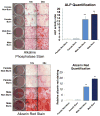
Results: Mesenchymal stem cells of male mice demonstrated greater osteogenic gene and protein expression than those of female mice (p < 0.05). Male mice in the burn group formed 35 percent more bone than female mice in the burn group. This bone formation correlated with increased pSmad and insulin-like growth factor 1 signaling at the heterotopic ossification site in male mice.
Conclusions: The authors demonstrate that male mice form quantitatively more bone compared with female mice using their burn/tenotomy model. These findings can be explained at least in part by differences in bone morphogenetic protein and insulin-like growth factor 1 signaling.
Conflict of interest statement
Disclosure Statement: The authors above have no financial interest in any of the products, devices, procedures or anything else connected with the article.
Figures


References
-
- Douglas K, Cannada LK, Archer KR, Dean DB, Lee S, Obremskey W. Incidence and risk factors of heterotopic ossification following major elbow trauma. Orthopedics. 2012;35(6):460. - PubMed
-
- Yi S, Shin DA, Kim KN, Choi G, Shin HC, Kim KS, Yoon DH. The predisposing factors for the heterotopic ossification after cervical artificial disc replacement. The Spine Journal. 2013;13(9):1048–1054. - PubMed
-
- Tu TH, Wu JC, Huang WC, Guo WY, Wu CL, Shih YH, Cheng H. Heterotopic ossification after cervical total disc replacement: determination by CT and effects on clinical outcomes: Clinical article. Journal of Neurosurgery: Spine. 2011;14(4):457–465. - PubMed
-
- Pavlou G, Salhab M, Murugesan L, Jallad S, Petsatodis G, West R, Tsiridis E. Risk factors for heterotopic ossification in primary total hip arthroplasty. Hip international: the journal of clinical and experimental research on hip pathology and therapy. 2011;22(1):50–55. - PubMed
-
- Medina A, Shankowsky H, Savaryn B, Shukalak B, Tredget E. Characterization of Heterotopic Ossification in Burn Patients. Journal of Burn Care and Research. 2013:1–6. - PubMed
Publication types
MeSH terms
Substances
Grants and funding
LinkOut - more resources
Full Text Sources
Medical

