. 2015 May 26;33(4):469-77.
doi: 10.1016/j.devcel.2015.03.018.

CXCL12 Signaling Is Essential for Maturation of the Ventricular Coronary Endothelial Plexus and Establishment of Functional Coronary Circulation

Affiliations

CXCL12 Signaling Is Essential for Maturation of the Ventricular Coronary Endothelial Plexus and Establishment of Functional Coronary Circulation

Susana Cavallero et al. Dev Cell. .

Abstract

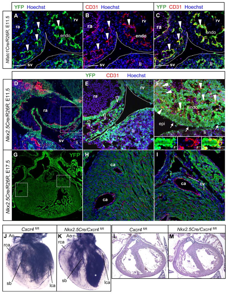
Maturation of a vascular plexus is a critical and yet incompletely understood process in organ development, and known maturation factors act universally in all vascular beds. In this study, we show that CXCL12 is an organ-specific maturation factor of particular relevance in coronary arterial vasculature. In vitro, CXCL12 does not influence nascent vessel formation, but promotes higher-order complexity of preinitiated vessels. In the heart, CXCL12 is expressed principally by the epicardium, and its receptor CXCR4 is expressed by coronary endothelial cells. CXCL12 is not a chemotactic signal for endothelial cell migration, but rather acts in a paracrine manner to influence the maturation of the coronary vascular plexus. Mutants in CXCL12 signaling show an excess of immature capillary chains and a selective failure in arterial maturation, and become leaky with the onset of coronary perfusion. Failed maturation of the coronary system explains the late-gestation lethality of these mutants.


Figures

Figure 1
Figure 1. CXCL12 promotes plexus maturation in vitro
(A–L) Representative images of HUVEC-covered beads in fibrin gels after 3, 5 and 7 days of culture. Negative control was endothelial basal media (EBM-2) plus 2% FBS with no added growth factors; VEGF and CXCL12 were added at 20 ng/ml and 100 ng/ml respectively. (M) Quantitation of total number of sprouts, tubes, branching points, and anastomosis in the fibrin bead assay. The data are normalized to the number of beads present in each microscopic field. Data are represented as mean ± SEM. * p<0.001 versus VEGF alone. See also Fig. S1.
Figure 2
Figure 2. CXCL12 and CXCR4 expression during coronary vessel development
(A–F) In situ hybridization with antisense probe for Cxcl12 shows expression in the epicardium in wild-type embryos. (A) Cxcl12 is expressed in the epicardium surrounding the atrioventricular canal at E10.5. (B) Hybridization with a sense control. A′ and B′ are magnified views of the boxed region in A and B. (C–F) Cxcl12 is expressed in the epicardium through E11.5–16.5, and in epicardium-derived smooth muscle surrounding coronary blood vessels (arrowheads) at E16.5. (G–I) CXCR4 is expressed in endothelial cells sprouting from the sinus venosus at E11.5 (arrowheads). The dotted lines outline the pericardial cavity. CD31 (Pecam1) is a pan-endothelial marker. Abbreviations: oft: outflow tract, ra: right atria, rv: right ventricle, avc: atrioventricular canal, sv, sinus venosus, li: liver, epi: epicardium. See also Fig. S2.
Figure 3
Figure 3. Defective coronary vessel organization in embryos with disrupted CXCL12 signaling
(A–H) Whole mount CD31 immunostaining of hearts (dorsal surfaces) from control and conditional Tie2Cre/Cxcr4 mutants at the indicated stages. Dotted lines indicate the extent of endothelial cell plexus organization within the ventricle. The diffuse staining at E11.5 on the ventricular surface corresponds to CD31-positive endocardium (open arrowheads). Views of the ventral surfaces of these same hearts are in Fig. S3. (I–L) Histological sections of control and mutant hearts at E13.5 immunostained with CD31 and endomucin (EMCN, venous marker). In the control, superficial veins are EMCN-positive (arrows). In the mutant, open arrowhead in center points to an intramyocardial vessel ectopically expressing endomucin, and the open arrowhead at right to a superficial dilated EMCN-negative vessel. (M–N) Reduced number of major coronary artery branches (SM22+) in mutant hearts at E15.5. (O–P) Whole mount EMCN staining at E15.5, indicating normal assembly of higher order venous structures (arrowheads). (Q–T) Visualization of the coronary arterial vasculature by aortic ink perfusion at E15.5 and E16.5. Arrowheads point to perfused vessels of abnormal branching organization in the mutant hearts. (U–V) Reduced number of major coronary artery branches in mutant hearts. U and V are low magnification views of control and mutant hearts at E17.5 showing only SM22 (smooth muscle marker) staining. Close-up views of the boxed regions are shown in U′-U″ and V′-V″ with CD31 and SM22 immunostaining. Endothelial cell density is similar between control and mutant (compare U′ to V′). Note smaller and fewer intramyocardial major branches (solid arrowheads) and dilated superficial vessels (open arrowheads) in the mutant. (W–X) Close up views of hearts from control (same as in Q) and Cxcl12 KO (shown in Fig. S4C). In the mutant, note perfusion of the proximal coronary artery from the aorta into the adjacent persisting capillary plexus. Abbreviations: Ao, aorta, endo, endocardium, lca, left coronary artery, rca, right coronary artery, sb, septal branch, co, coronary ostia. See also Fig. S3.
Figure 4
Figure 4. Coronary endothelial cells are derived from the sinus venosus
(A–C) Nfatc1Cre recombines in sinus venosus endothelial cell sprouts (three arrowheads at left) and in all endocardial cells at E11.5. (D–E) Nkx2.5Cre recombination at E11.5. Panel E corresponds to the boxed area in D. Nkx2.5Cre does not recombine in endothelial cells (CD31+) of the sinus venosus and in endothelial cell sprouts of the sinus venosus moving towards the ventricle at the atrioventricular groove (open arrowheads). (F) Nkx2.5Cre recombines efficiently in the myocardium, epicardium (arrows) and the majority of endocardial cells (solid arrowheads). A close-up confocal view of a trabecular element is shown in separate (F′ and F″) and merged (F‴) channels in the insets to emphasize that endocardial cells are labeled. (G–I) Nkx2.5Cre recombination at E17.5. Panel G shows a low magnification view of the heart (green channel only). H and I correspond to the boxed areas in G. Nkx2.5Cre does not recombine in coronary artery or vein endothelium throughout the mature ventricle. (J–K) Normal coronary vasculature in E17.5 Nkx2.5Cre/Cxcr4 mutants; the asterisk in K indicates leaking of excess ink into the ventricular cavity. (L–M) H&E staining of frontal sections showing normal heart morphology in both control and mutant hearts at E17.5. Abbreviations: ra, right atria; sv, sinus venosus, rv, right ventricle, endo, endocardium, li, liver; ca, coronary artery, cv, coronary vein, lca, left coronary artery, rca, right coronary artery, sb, septal branch. See also Fig. S4.

References

    1. Ara T, Nakamura Y, Egawa T, Sugiyama T, Abe K, Kishimoto T, Matsui Y, Nagasawa T. Impaired colonization of the gonads by primordial germ cells in mice lacking a chemokine, stromal cell-derived factor-1 (SDF-1) Proc Natl Acad Sci U S A. 2003;100:5319–5323. - PMC - PubMed
    1. Ara T, Tokoyoda K, Okamoto R, Koni PA, Nagasawa T. The role of CXCL12 in the organ-specific process of artery formation. Blood. 2005;105:3155–3161. - PubMed
    1. Carmeliet P, Ferreira V, Breier G, Pollefeyt S, Kieckens L, Gertsenstein M, Fahrig M, Vandenhoeck A, Harpal K, Eberhardt C, et al. Abnormal blood vessel development and lethality in embryos lacking a single VEGF allele. Nature. 1996;380:435–439. - PubMed
    1. Chen HI, Poduri A, Numi H, Kivela R, Saharinen P, McKay AS, Raftrey B, Churko J, Tian X, Zhou B, et al. VEGF-C and aortic cardiomyocytes guide coronary artery stem development. J Clin Invest. 2014;124:4899–4914. - PMC - PubMed
    1. Choudhary B, Zhou J, Li P, Thomas S, Kaartinen V, Sucov HM. Absence of TGFbeta signaling in embryonic vascular smooth muscle leads to reduced lysyl oxidase expression, impaired elastogenesis, and aneurysm. Genesis. 2009;47:115–121. - PubMed

Publication types

MeSH terms

LinkOut - more resources