. 2015 Aug;89(15):7979-90.
doi: 10.1128/JVI.00375-15. Epub 2015 May 27.

Promotion of Endoplasmic Reticulum-Associated Degradation of Procathepsin D by Human Herpesvirus 8-Encoded Viral Interleukin-6

Affiliations

Promotion of Endoplasmic Reticulum-Associated Degradation of Procathepsin D by Human Herpesvirus 8-Encoded Viral Interleukin-6

Daming Chen et al. J Virol. 2015 Aug.

Abstract

The interleukin-6 homologue (viral interleukin-6 [vIL-6]) of human herpesvirus 8 is implicated in viral pathogenesis due to its proproliferative, inflammatory, and angiogenic properties, effected through gp130 receptor signaling. In primary effusion lymphoma (PEL) cells, vIL-6 is expressed latently and is essential for normal cell growth and viability. This is mediated partly via suppression of proapoptotic cathepsin D (CatD) via cocomplexing of the endoplasmic reticulum (ER)-localized CatD precursor, pro-CatD (pCatD), and vIL-6 with the previously uncharacterized ER membrane protein vitamin K epoxide reductase complex subunit 1 variant 2 (VKORC1v2). vIL-6 suppression of CatD occurs also during reactivated productive replication in PEL cells and is likely to contribute to proreplication functions of vIL-6. Here, we report that vIL-6 suppresses CatD through vIL-6, VKORC1v2, and pCatD association with components of the ER-associated degradation (ERAD) machinery. In transfected cells, expression of vIL-6 along with CatD led to proteasome-dependent (inhibitor-sensitive) decreases in CatD levels and the promotion of pCatD polyubiquitination. Depletion of particular ERAD-associated isomerases, lectins, and translocon components, including ERAD E3 ubiquitin ligase HRD1, diminished suppression of CatD by vIL-6. Coprecipitation assays identified direct or indirect interactions of VKORC1v2, vIL-6, and pCatD with translocon proteins (SEL1L and/or HRD1) and ERAD-associated lectins OS9 and XTP3-B. Endogenous CatD expression in PEL cells was increased by depletion of ERAD components, and suppression of CatD by vIL-6 overexpression in PEL cells was dependent on HRD1. Our data reveal a new mechanism of ER-localized vIL-6 activity and further characterize VKORC1v2 function.

Importance: Human herpesvirus 8 (HHV-8) viral interleukin-6 (vIL-6), unlike cellular IL-6 proteins, is secreted inefficiently and sequestered mainly in the endoplasmic reticulum (ER), from where it can signal through the gp130 receptor. We have recently reported that vIL-6 also associates with a novel membrane protein termed vitamin K epoxide reductase complex subunit 1 variant 2 (VKORC1v2) and mediates suppression of VKORC1v2-cointeracting cathepsin D, a stress-released proapoptotic protein negatively impacting HHV-8 latently infected primary effusion lymphoma (PEL) cell viability and reactivated virus productive replication. Here, we have examined the mechanistic basis of the VKORC1v2-vIL-6 interaction-dependent suppression of cathepsin D and have found that this novel activity of vIL-6 is mediated through coassociation of VKORC1v2, procathepsin D, and vIL-6 with components of the ER-associated degradation (ERAD) machinery. Our findings provide information of significance for potential antiviral and therapeutic targeting of VKORC1v2-mediated vIL-6 activities and also indicate the nature of VKORC1v2 function in normal cell biology.


Figures

FIG 1
FIG 1
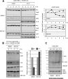
vIL-6-regulated expression of pCatD. (A) HEK293T cells were cotransfected with expression vectors for CatD and either empty vector (vec) or increasing amounts of vIL-6-expressing plasmid (0.1 to 0.8 μg), either in the absence (left panels) or in the presence (right panels) of VKORC1v2 (v2) vector. CatD and YFP contiguous coding sequences were linked by those encoding porcine teschovirus peptide 2A (P2A) in the CatD expression vector to allow single-transcript cotranslation but separate expression (illustrated). CatD levels in cell lysates and culture media harvested 24 h posttransfection were determined by Western blotting; for the latter, pCatD was first concentrated by pepstatin A bead precipitation (see Materials and Methods). ER-localized pro-CatD (p) and lysosomal mature CatD (m) are indicated, along with likely unglycosylated pCatD (*). (B) Quantified immunoblot data, showing the vIL-6-influenced levels of intracellular pCatD and mCatD in relation to levels in the presence of empty vector. Values were normalized to β-actin.
FIG 2
FIG 2
Proteasome involvement in CatD regulation by vIL-6. (A) KDEL motif-tagged (ER-retained) pCatD was expressed along with VKORC1v2 in transfected HEK293T cells, either without (vec) or with vIL-6 coexpression. At 24 h posttransfection, cultures were treated with cycloheximide (Cx; 100 μg/ml) to block translation and either proteasome inhibitor MG132 (10 μM) or DMSO (vehicle; control). Cells were harvested 0 to 8 h after drug addition for immunoblot analysis of cell lysates. pCatD (p) and mature (m) endogenous CatD are indicated. Quantified data (right) show β-actin-normalized pCatD levels after cycloheximide treatment relative to time zero levels (set at 1). (B) An analogous experiment was undertaken, but employing an expression vector for P2A-linked pCatD-KDEL and YFP. Cells were harvested either at initiation or after 7 h of cycloheximide and MG132 or DMSO treatment. The graph shows actin-normalized levels of pCatD-KDEL expression at 7 h after cycloheximide treatment relative to time zero levels (set at 1). (C) HEK293T cultures cotransfected with expression plasmids for CatD-CBD and vIL-6 or empty vector (vec) were treated for 6 h with DMSO (control) or MG132 (10 μM). Chitin bead-precipitated material (normalized for pCatD) was analyzed by Western blotting for detection of CatD and ubiquitin. Mono- and polyubiquitinated species are indicated.
FIG 3
FIG 3
ERAD protein involvement in vIL-6 suppression of CatD. (A) Illustration of ERAD components and associated lectins, chaperones, and protein disulfide isomerases and oxidoreductases. The glycoprotein-binding ERAD lectins interact with the translocon complex, which includes Derlin, SEL1L, and HRD1 (shown), in addition to other proteins, to enable retrotranslocation of unfolded and ultimately deglycosylated proteins (19, 21). PDI, protein disulfide isomerase. (B) Validation of shRNAs reported previously to be active in HEK293T cells (19) was undertaken by cotransfection of effector (+) or nonsilencing (NS) control (−) shRNA expression vectors, either without (Derlin-1 and ERdj5) or with plasmids expressing S-epitope-tagged target proteins. Immunoblotting or RT-PCR was used for detection of proteins or transcripts. Glyceraldehyde-3-phosphate dehydrogenase (GAPDH)-specific RT-PCR provided a normalization control; no PCR products were detected absent reverse transcription (data not shown). (C) HEK293T cells transduced with lentiviral vectors expressing ERAD mRNA-targeting or control (NS) shRNAs were transfected with YFP-P2A-CatD expression vector and either a vIL-6-encoding or empty vector (vec). Cells were harvested 24 h posttransfection, and lysates were analyzed by Western blotting. The graph (right) shows YFP-normalized expression levels of pCatD (upper band) in vIL-6-expressing cultures relative to empty-vector-transfected cultures.
FIG 4
FIG 4
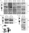
VKORC1v2 interactions with ERAD-associated proteins. (A) HEK293T cells were cotransfected with vectors encoding VKORC1v2-CBD (or VKORC1v1-CBD, control), CatD, and vIL-6 (or empty vector, “vec”) together with plasmids specifying S-tagged SEL1L, OS9, XTP3-B, HRD1, or gp78. Cell lysates and chitin bead-precipitated proteins were gel fractionated and immunoblotted for detection of expression and coprecipitation of S tag (ERAD proteins), CatD, and vIL-6. (B) Transfections and analyses for SEL1L-S and HRD1-S were as outlined for panel A except that CatD expression vector was not transfected and no CatD immunoblotting was done (endogenous pCatD is present at very low levels [9]). (C) A similar experiment was undertaken with and without CatD vector cotransfection to examine the potential influence of pCatD on VKORC1v2-HRD1 association. (D) Diagrammatic summary of data, indicating (direct or indirect) interactions of precipitated VKORC1v2 (shown in black) with ERAD lectins OS9 and XTP3-B and translocon components SEL1L and HRD1. Dotted arrows indicate hypothesized interactions of pCatD with the lectins prior to and/or (?) after their association with the translocon. Promotion of pCatD-VKORC1v2 association by vIL-6, published previously (9) and confirmed here, is indicated by the arrow and plus sign.
FIG 5
FIG 5
vIL-6 association with ERAD proteins. (A) S-epitope-tagged SEL1L, OS9, XTP3-B, and HRD1 (or empty vector control) were each expressed along with CatD and Twin-StrepII (TS)-tagged vIL-6 (or empty vector [vec]) and either (CBD-fused) VKORC1v2 (v2) or vIL-6-refractory VKORC1v2.ΔvBD (v2Δ), deleted of the vIL-6-binding domain (16). StrepTactin bead-sedimented material from transfected HEK293T cell lysates was gel fractionated and immunoblotted for detection of vIL-6-TS-coprecipitated proteins. (B) Illustrated summary of the data, indicating direct or indirect interactions of vIL-6 with lectins OS9 and XTP3-B and with translocon E3 ligase HRD1. Asterisks indicate VKORC1v2-independent (vBD mutation-refractory) associations of vIL-6 with the lectins and also with pCatD specifically in XTP3-B vector-transfected cells. Dashed lines indicate lectin interactions with vIL-6 and/or CatD either separately or as part of the translocon complex (undetermined). Expected pCatD-lectin interactions with the translocon may or may not involve VKORC1v2-associated pCatD (?).
FIG 6
FIG 6
Interactions of pCatD with ERAD components. (A) CBD-fused SEL1L was used as “bait” in a coprecipitation experiment to assay for SEL1L interactions with CatD and vIL-6. S-tagged HRD1 was used as a positive control for interaction with SEL1L; empty vector (vec) was used as a negative control. (B) S-tagged ERAD lectins OS9 and XTP3-B were expressed at different doses (0.5 to 10 μg vectors) along with CatD and vIL-6 in appropriately transfected HEK293T cells for S-peptide-based coprecipitation analysis of pCatD-lectin binding and vIL-6 association. Empty vector (vec) was used as a negative control and filler. (C) Illustration and modeling of the findings from experiments shown in panels A and B, indicating translocon-independent and -associated complexing of pCatD with lectins OS9 and XPT3-B and detected (HRD1-independent) SEL1L interactions with pCatD and vIL-6 [with (?) or without VKORC1v2 involvement].
FIG 7
FIG 7
Operation of the ERAD pathway in CatD regulation in PEL cells. (A) BCBL-1 cultures were transduced via lentiviral vector infection with shRNAs targeting transcripts of translocon proteins SEL1L, HRD1, and Derlin-1 or with nonsilencing (NS) control shRNA. For validation of shRNA activities, semiquantitative PCR was applied to reverse-transcribed mRNA samples from transduced cultures to detect shRNA target and control glyceraldehyde-3-phosphate dehydrogenase (GAPDH) mRNA. No PCR products were detected in non-reverse-transcribed samples (data not shown). (B) Western blot analysis of CatD expression in equivalently transduced cultures. CatD levels in cell lysates relative to β-actin are shown as CatD/actin ratios below the respective lanes (“NS” value set at 1.0). (C) A similar experiment was undertaken in which SEL1L or HRD1 was depleted either alone (with NS [control] shRNA cotransduction) or together with vIL-6 codepletion. The relative levels of actin-normalized CatD (CatD/actin, “double NS” shRNA transduction set at 1.0) are indicated below the respective lanes. The graph (right) quantifies the vIL-6 depletion-mediated CatD induction in the absence (NS) and presence of translocon protein depletion. (D) BCBL-1 cells were cotransduced with empty (vec) or vIL-6-expressing lentiviral vector and either HRD1 transcript-targeting or NS shRNA-expressing lentivirus. Levels of CatD relative to β-actin are shown below the respective immunoblot lanes (“NS + vec” set at 1). The graph (right) displays the relative levels of vIL-6-mediated CatD suppression in the absence and presence of HRD1 depletion.
FIG 8
FIG 8
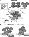
Overview of main findings and associated models. (A) Modeling of combined coprecipitation and functional data, indicating (i) detected (pCatD-independent) interactions between VKORC1v2 and translocon components SEL1L and HRD1 in addition to lectins OS9 and XTP3-B (from Fig. 4); (ii) vIL-6-promoted pCatD-VKORC1v2 association (Fig. 4) (9); (iii) VKORC1v2-independent vIL-6–lectin and vIL-6–pCatD (XTP3-B-involved) interactions (Fig. 5); (iv) pCatD-translocon and -lectin interactions (Fig. 6); and (v) functional involvement in vIL-6-mediated CatD suppression of ERAD translocon, lectin, chaperone, and cochaperone proteins (boxed), as revealed by depletion experiments (Fig. 3 and 7). It is speculated that vIL-6 can promote pCatD-ERAD association, and consequent pCatD degradation, via stabilization of pCatD binding to translocon-associated VKORC1v2 and possibly also by vIL-6–VKORC1v2 interaction-mediated recruitment of pCatD-lectin complexes to the translocon. (B) Cocomplexes of vIL-6, XTP3-B, and pCatD, formed independently of VKORC1v2, could be important for delivery of pCatD to the ERAD complex, either containing functionally involved OS9 or independently of the lectin.

References

    1. Arvanitakis L, Mesri EA, Nador RG, Said JW, Asch AS, Knowles DM, Cesarman E. 1996. Establishment and characterization of a primary effusion (body cavity-based) lymphoma cell line (BC-3) harboring Kaposi's sarcoma-associated herpesvirus (KSHV/HHV-8) in the absence of Epstein-Barr virus. Blood 88:2648–2654. - PubMed
    1. Carbone A, Cilia AM, Gloghini A, Capello D, Perin T, Bontempo D, Canzonieri V, Tirelli U, Volpe R, Gaidano G. 2000. Primary effusion lymphoma cell lines harbouring human herpesvirus type-8. Leuk Lymphoma 36:447–456. doi:10.3109/10428190009148391. - DOI - PubMed
    1. Chang Y, Moore PS. 1996. Kaposi's sarcoma (KS)-associated herpesvirus and its role in KS. Infect Agents Dis 5:215–222. - PubMed
    1. Gaidano G, Pastore C, Gloghini A, Volpe G, Capello D, Polito P, Vaccher E, Tirelli U, Saglio G, Carbone A. 1997. Human herpesvirus type-8 (HHV-8) in haematopoietic neoplasia. Leuk Lymphoma 24:257–266. - PubMed
    1. Chen D, Sandford G, Nicholas J. 2009. Intracellular signaling mechanisms and activities of human herpesvirus 8 interleukin-6. J Virol 83:722–733. doi:10.1128/JVI.01517-08. - DOI - PMC - PubMed

Publication types

MeSH terms

LinkOut - more resources