. 2015 Aug;89(16):8258-66.
doi: 10.1128/JVI.01045-15. Epub 2015 May 27.

Functional Analysis of the 60-Nucleotide Duplication in the Respiratory Syncytial Virus Buenos Aires Strain Attachment Glycoprotein

Affiliations

Functional Analysis of the 60-Nucleotide Duplication in the Respiratory Syncytial Virus Buenos Aires Strain Attachment Glycoprotein

Anne L Hotard et al. J Virol. 2015 Aug.

Abstract

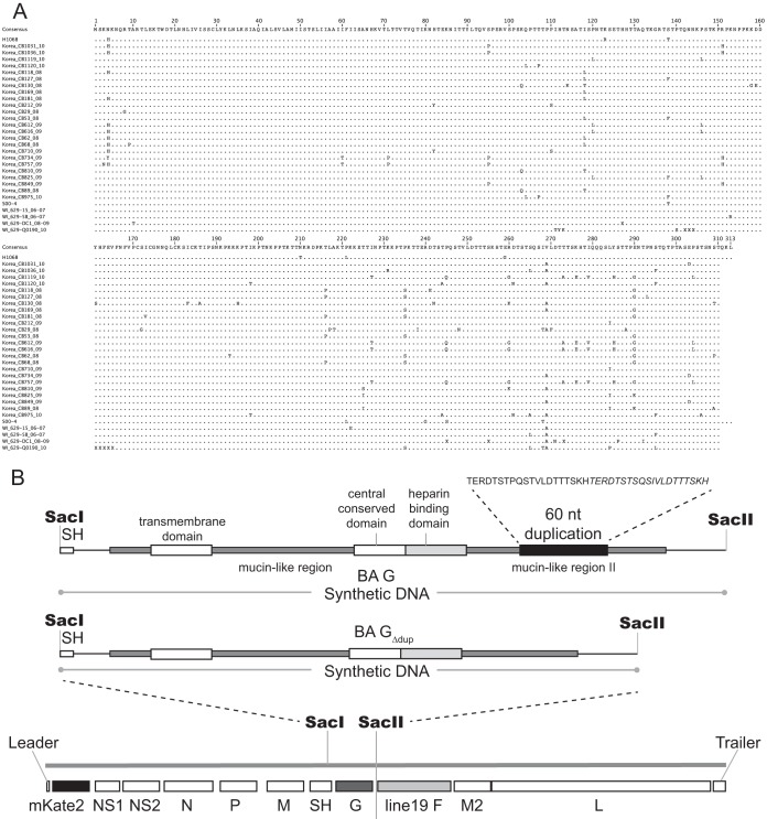
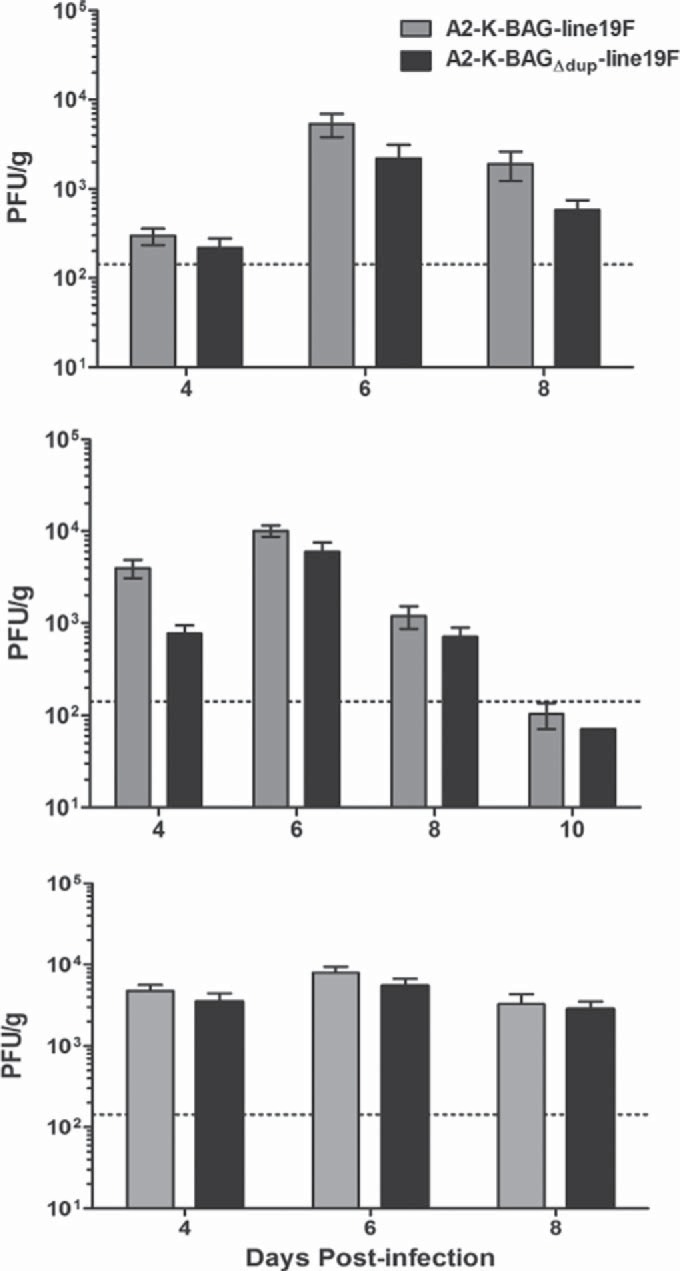
There are two subgroups of respiratory syncytial virus (RSV), A and B, and within each subgroup, isolates are further divided into clades. Several years ago, multiple subgroup B isolates which contained a duplication of 60 nucleotides in the glycoprotein (G) gene were described. These isolates were given a new clade designation of BA based on the site of isolation, Buenos Aires, Argentina. BA RSV strains have since become the predominant circulating clade of RSV B viruses. We hypothesized that the duplicated region in G serves to enhance the function of G in the virus life cycle. We generated recombinant viruses that express a consensus BA G gene or a consensus BA G gene lacking the duplication (GΔdup). We determined that the duplicated region functions during virus attachment to cells. Additionally, we showed that in vitro, the virus containing the duplication has a fitness advantage compared to the virus without the duplication. Our data demonstrate that the duplicated region in the BA strain G protein augments virus attachment and fitness.

Importance: Respiratory syncytial virus (RSV) is an important pathogen for infants for which there is no vaccine. Different strains of RSV circulate from year to year, and the predominating strains change over time. Subgroup B RSV strains with a duplication in the attachment glycoprotein (G) emerged and then became the dominant B genotype. We found that a recombinant virus harboring the duplication bound more efficiently to cells and was more fit than a recombinant strain lacking the duplication. Our work advances a mechanism for an important natural RSV mutation.


Figures

FIG 1
FIG 1
Recombinant virus design. (A) Amino acid alignment used to generate the consensus sequences. Numbering is based on the consensus. Amino acid disagreements from the consensus are indicated. The final QKL residues were not included in the consensus used for cloning. The duplication spans residues 258 to 277. (B) Subclones harboring the designed consensus sequences of BA G or BA GΔdup were obtained from GeneArt. SacI and SacII restriction sites flanking the G genes were utilized to clone the G constructs into pSynkRSVline19F in place of A2 G. Important domains of G are labeled, and the amino acid sequence of the duplication is shown, with the initial run of amino acids in regular type and the inexact duplication in italics.
FIG 2
FIG 2
Glycoprotein incorporation into recombinant viruses. Virus stocks of each recombinant virus were purified through a 20% sucrose cushion and subjected to SDS-PAGE and Western blotting for the G or F glycoproteins or the RSV nucleoprotein as a loading control. Images shown are from Western blot analyses performed on a single membrane probed for each protein sequentially and represent results from the purification of two independent sets of virus stocks. Molecular masses of marker bands are shown in kilodaltons.
FIG 3
FIG 3
Recombinant BA strain replication in vitro. (A) BEAS-2B cells were infected with the two viruses indicated at an MOI of 0.01. Cells were scraped into medium at the indicated time points, and virus was titrated by a fluorescent focus-forming assay in HEp-2 cells. The data are a combination of three experiments. There were no significant differences based on a two-way analysis of variance (ANOVA). (B) Single vials of virus stocks were titrated in both CHO-K1 and pgsD-677 cell lines. Each experiment was done in triplicate, and the graph shows a combination of results of three experiments. *, P < 0.05 (by one-way ANOVA and Tukey's multiple-comparison test). n.s., not significant.
FIG 4
FIG 4
Recombinant BA strain binding to cells in vitro. (A and B) BEAS-2B cells were inoculated with the indicated viruses at an MOI of 1.0, and virus was allowed to adsorb to cells at 4°C. Inoculum was removed, and cells were washed three times in cold PBS to remove unbound virus. Cells were lysed, and lysates, along with an aliquot of original inoculum, was subjected to SDS-PAGE and Western blotting with an anti-N monoclonal antibody. GAPDH was also probed as a loading control. (A) Representative Western blots of binding assay. (B) Combination of densitometry results of the three experiments illustrated in panel A. The amount of bound N was normalized to N in the inoculum as well as GAPDH prior to comparison between groups. *, P < 0.05 (by paired t test). (C) Representative Western blots from binding assay performed with CHO-K1 and pgsD-677 cell lines. For experiments in CHO-K1 and pgsD-677 cells, sucrose-purified virus stocks were used, and the infection inoculum was normalized based on N protein levels in purified stocks. (D) Combination of densitometry results of the three experiments illustrated in panel C. *, P < 0.05 (by paired t test). ns, not significant.
FIG 5
FIG 5
Competitive-infection assay. HEp-2 cells were infected with A2-K-BAG-line19F, A2-K-BAGΔdup-line19F, or both viruses at a ratio of 1:1, 10:1, or 1:10. Twenty-four and 48 h postinfection, RNA was harvested from cells and subjected to reverse transcription (RT)-PCR to amplify a portion of the G gene. Equal amounts of RNA were used for the reverse transcription, and equal volumes of cDNA were used as templates for the PCR. Gels are representative of two experiments. Expected PCR product sizes are 709 bp for A2-K-BAG-line19F and 649 bp for A2-K-BAGΔdup-line19F.
FIG 6
FIG 6
Recombinant BA strain lung viral load in BALB/c mice. Groups of 5 BALB/c mice were infected with 1 × 10⁵ PFU of either A2-K-BAG-line19F or A2-K-BAGΔdup-line19F, and lungs were harvested on day 4, 6, 8, or 10 postinfection. Viral load was quantified by an immunodetection plaque assay. Each graph is an independent experiment. There were no significant differences. A horizontal dotted line indicates the limit of detection.
FIG 7
FIG 7
Microneutralization assay of recombinant BA viruses. A2-K-BAG-line19F and A2-K-BAGΔdup-line19F were incubated with PBS or with serial dilutions of motavizumab, an F-specific monoclonal antibody, and then used to infect HEp-2 cells. The level of neutralization of each virus was calculated by comparing the number of FFU present when the virus was mixed with antibody dilutions to the number of FFU present when the virus was mixed with PBS. The results shown are representative of three replicate experiments.

References

    1. Collins PL, Crowe JE Jr. 2007. Respiratory syncytial virus and metapneumovirus, p 1601–1646. In Knipe DM, Howley PM (ed), Fields virology, vol 2 Lippincott, Williams, and Wilkins, Philadelphia, PA.
    1. Peret TC, Hall CB, Schnabel KC, Golub JA, Anderson LJ. 1998. Circulation patterns of genetically distinct group A and B strains of human respiratory syncytial virus in a community. J Gen Virol 79(Pt 9):2221–2229. - PubMed
    1. Trento A, Galiano M, Videla C, Carballal G, Garcia-Barreno B, Melero JA, Palomo C. 2003. Major changes in the G protein of human respiratory syncytial virus isolates introduced by a duplication of 60 nucleotides. J Gen Virol 84:3115–3120. doi:10.1099/vir.0.19357-0. - DOI - PubMed
    1. Trento A, Viegas M, Galiano M, Videla C, Carballal G, Mistchenko AS, Melero JA. 2006. Natural history of human respiratory syncytial virus inferred from phylogenetic analysis of the attachment (G) glycoprotein with a 60-nucleotide duplication. J Virol 80:975–984. doi:10.1128/JVI.80.2.975-984.2006. - DOI - PMC - PubMed
    1. Trento A, Casas I, Calderon A, Garcia-Garcia ML, Calvo C, Perez-Brena P, Melero JA. 2010. Ten years of global evolution of the human respiratory syncytial virus BA genotype with a 60-nucleotide duplication in the G protein gene. J Virol 84:7500–7512. doi:10.1128/JVI.00345-10. - DOI - PMC - PubMed

Publication types

MeSH terms

LinkOut - more resources