Do Mesenchymal Stromal Cells Influence Microscopic Residual or Metastatic Osteosarcoma in a Murine Model?
- PMID: 26018200
- PMCID: PMC4746149
- DOI: 10.1007/s11999-015-4362-2
Do Mesenchymal Stromal Cells Influence Microscopic Residual or Metastatic Osteosarcoma in a Murine Model?
Abstract
Background: Mesenchymal stromal cells (MSCs) have been shown in rodent models to promote primary and pulmonary metastatic sarcoma growth when injected in the presence of gross tumor. In theory, this would limit their use in a clinical setting after limb salvage treatment for osteosarcoma. Although concerning, these models do not translate to the clinical setting wherein MSCs could be used after primary tumor resection to aid in bone healing and incorporation of tumor endoprostheses. If we can determine whether the use of MSCs in this setting is safe, it might improve our ability to augment bone healing in patients undergoing limb salvage.
Questions/purposes: The purpose of this study was to determine (1) whether MSCs promote pulmonary metastatic disease progression in a murine osteosarcoma model; and/or (2) whether they affect local disease recurrence in the presence of microscopic residual osteosarcoma.
Methods: An orthotopic model of luciferase-expressing osteosarcoma was developed. At 10 days, resection of the primary tumor was performed. One hundred fourteen female C3H mice were inoculated with DLM8-luc osteosarcoma in the proximal tibia. Ninety-four mice developed orthotopic osteosarcoma with luciferase expression. Mice with bioluminescent evidence of a primary tumor received either a microscopically "clean" amputation at a time when residual microscopic metastatic disease was present in the lungs (pulmonary metastasis group; n = 65) or a "dirty" amputation (local recurrence group; n = 29). Mice were randomized to receive intravenous MSCs, MSCs at the surgical site, or no MSCs. Mice were monitored for development and progression of pulmonary metastasis and local recurrence by bioluminescence imaging and daily measurements at the surgical site. The number of pulmonary nodules, time to first evidence of metastasis, and size of recurrent tumor were compared using Kruskal-Wallis, analysis of variance, Welch's, t-tests, or Mann-Whitney tests as appropriate for the specific data sets with p < 0.05 considered significant.
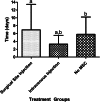
Results: Mice receiving intravenous MSCs had a faster time to first detection of pulmonary metastasis (2.93 ± 1.90 days) compared with mice with local injection of MSCs (6.94 ± 6.78 days) or no MSCs (5.93 ± 4.55 days) (p = 0.022). MSC treatment did not influence whether mice developed local recurrence (p = 0.749) or size of recurrent tumors (p = 0.221).
Conclusions: MSCs delivered to the surgical site did not promote local recurrence or size of recurrent tumors, but intravenous injection of MSCs did hasten onset of detection of pulmonary metastatic disease. Although local administration of MSCs into a surgical site does not appear to promote either pulmonary metastatic disease or local recurrence, large variation within groups and small numbers diminished statistical power such that a Type II error cannot be ruled out.
Clinical relevance: If MSCs are to be used to augment bone healing in the postlimb salvage setting in patients with osteosarcoma, it will be important to understand their influence, if any, on pulmonary micrometastsis or residual microscopic local disease. Although murine models do not completely recapitulate the clinical scenario, these results suggest that intravenous delivery of MSCs may promote micrometastatic pulmonary disease. Local administration into a surgical wound, even in the presence of residual microscopic disease, may be safe, at least in this murine model, but further investigation is warranted before considering the use of MSCs for clinical use in patients with osteosarcoma.
Figures


Comment in
-
CORR Insights(®): Do Mesenchymal Stromal Cells Influence Microscopic Residual or Metastatic Osteosarcoma in a Murine Model?Clin Orthop Relat Res. 2016 Mar;474(3):716-8. doi: 10.1007/s11999-015-4423-6. Epub 2015 Jun 24. Clin Orthop Relat Res. 2016. PMID: 26105153 Free PMC article. No abstract available.
References
-
- Asai T, Ueda T, Itoh K, Yoshioka K, Aoki Y, Mori S, Yoshikawa H. Establishment and characterization of a murine osteosarcoma cell line (LM8) with high metastatic potential to the lung. Int J Cancer. 1998;76:418–422. doi: 10.1002/(SICI)1097-0215(19980504)76:3<418::AID-IJC21>3.0.CO;2-5. - DOI - PubMed
-
- Berlin O, Samid D, Donthineni-Rao R, Akeson W, Amiel D, Woods VL., Jr Development of a novel spontaneous metastasis model of human osteosarcoma transplanted orthotopically into bone of athymic mice. Cancer Res. 1993;53:4890–4895. - PubMed
MeSH terms
Grants and funding
LinkOut - more resources
Full Text Sources
Other Literature Sources
Medical
Research Materials

