. 2015 May 27;10(5):e0126352.
doi: 10.1371/journal.pone.0126352. eCollection 2015.

C-Terminal Clostridium perfringens Enterotoxin-Mediated Antigen Delivery for Nasal Pneumococcal Vaccine

Affiliations

C-Terminal Clostridium perfringens Enterotoxin-Mediated Antigen Delivery for Nasal Pneumococcal Vaccine

Hidehiko Suzuki et al. PLoS One. .

Abstract

Efficient vaccine delivery to mucosal tissues including mucosa-associated lymphoid tissues is essential for the development of mucosal vaccine. We previously reported that claudin-4 was highly expressed on the epithelium of nasopharynx-associated lymphoid tissue (NALT) and thus claudin-4-targeting using C-terminal fragment of Clostridium perfringens enterotoxin (C-CPE) effectively delivered fused antigen to NALT and consequently induced antigen-specific immune responses. In this study, we applied the C-CPE-based vaccine delivery system to develop a nasal pneumococcal vaccine. We fused C-CPE with pneumococcal surface protein A (PspA), an important antigen for the induction of protective immunity against Streptococcus pneumoniae infection, (PspA-C-CPE). PspA-C-CPE binds to claudin-4 and thus efficiently attaches to NALT epithelium, including antigen-sampling M cells. Nasal immunization with PspA-C-CPE induced PspA-specific IgG in the serum and bronchoalveolar lavage fluid (BALF) as well as IgA in the nasal wash and BALF. These immune responses were sufficient to protect against pneumococcal infection. These results suggest that C-CPE is an efficient vaccine delivery system for the development of nasal vaccines against pneumococcal infection.


Conflict of interest statement

Competing Interests: The authors have declared that no competing interests exist.

Figures

Fig 1
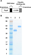
Fig 1. Construction and preparation of PspA-C-CPE.
(A) Schematic illustration of PspA-C-CPE. PspA was fused with C-CPE at its N-terminus. A G4S linker was inserted between PspA and C-CPE. (B) PspA and PspA-C-CPE were expressed in Escherichia coli as His-tagged proteins and purified by Ni-affinity chromatography. The PspA and PspA-C-CPE recombinant protein were applied to SDS-PAGE followed by staining with Coomassie brilliant blue. Lane 1, size ladder; lane 2, PspA; lane 3, PspA-C-CPE.
Fig 2
Fig 2. Binding of PspA-C-CPE to claudin-4-expressing cells.
(A) Claudin-4-expressing L cells and parent L cells were treated with PspA or PspA-C-CPE. Their bindings were detected by using an anti-His tag antibody followed by staining with fluorescein-labeled secondary antibody. Violet histograms are PspA or PspA-C-CPE; the green line histogram is control. (B) Binding of PspA-C-CPE to NALT epithelium. NALT sections were fixed with acetone and stained with biotinylated-PspA or biotinylated-PspA-C-CPE followed by staining with Alexa Fluor 546-conjugated streptavidin. M cells were detected by staining with fluorescein-conjugated UEA-1. Yellow arrows indicate PspA-C-CPE bound to M cells. Red, biotinylated-PspA or biotinylated-PspA-C-CPE; green, UEA-1; blue, DAPI. Scale bar is 100 μm (left) or 50 μm (right).
Fig 3
Fig 3. Induction of PspA-specific systemic and respiratory antibody responses by intranasal immunization with PspA-C-CPE.
Mice were nasally immunized with vehicle, PspA alone, or PspA-C-CPE (PspA; 5 μg) once weekly for 3 weeks. One week after the last immunization, PspA-specific serum IgG (A), nasal IgA (B), BALF IgG (C), and IgA (D) were measured by ELISA. Data are shown as mean ± SEM and are representative of two independent experiments. Vehicle, n = 4; PspA, n = 5; PspA-C-CPE, n = 5. Values were compared by using the non-parametric Mann–Whitney U test. *P < 0.01.
Fig 4
Fig 4. PspA-C-CPE-mediated induction of protective immunity against pneumococcal infection.
Mice were nasally immunized with vehicle, PspA alone, or PspA-C-CPE (PspA; 5μg) once weekly for 3 weeks. One week after the last immunization, mice were intrarespiratory challenged with S. pneumoniae (5.0 × 10⁶ CFU/mouse), and their survival was monitored for 14 days. Survival was compared between groups by using the non-parametric Mann–Whitney U test. **P < 0.05, *P < 0.01. Data were collected four experiments. Vehicle, n = 35; PspA, n = 45; PspA-C-CPE, n = 59.

References

    1. Fujkuyama Y, Tokuhara D, Kataoka K, Gilbert RS, McGhee JR, Yuki Y, et al. Novel vaccine development strategies for inducing mucosal immunity. Expert Review of Vaccines. 2012;11(3):367–79. 10.1586/erv.11.196 - DOI - PMC - PubMed
    1. Dlugonska H, Grzybowski M. Mucosal vaccination—an old but still vital strategy. Annals of Parasitology. 2012;58(1):1–8. - PubMed
    1. Mabbott NA, Donaldson DS, Ohno H, Williams IR, Mahajan A. Microfold (M) cells: important immunosurveillance posts in the intestinal epithelium. Mucosal Immunol. 2013;6(4):666–77. 10.1038/mi.2013.30 - DOI - PMC - PubMed
    1. Brandtzaeg P. Induction of secretory immunity and memory at mucosal surfaces. Vaccine. 2007;25(30):5467–84. - PubMed
    1. Kunisawa J, Kurashima Y, Kiyono H. Gut-associated lymphoid tissues for the development of oral vaccines. Advanced Drug Delivery Reviews. 2012;64(6):523–30. 10.1016/j.addr.2011.07.003 - DOI - PubMed

Publication types

MeSH terms