

Evaluation of protein undernourishment on the condylar process of the Wistar rat mandible correlation with insulin receptor expression
- PMID: 26018304
- PMCID: PMC4428457
- DOI: 10.1590/1678-775720140319
Evaluation of protein undernourishment on the condylar process of the Wistar rat mandible correlation with insulin receptor expression
Abstract
The mandible condylar process cartilage (CP) of Wistar rats is a secondary cartilage and acts as a mandibular growth site. This phenomenon depends on adequate proteins intake and hormone actions, including insulin.
Objectives: The present study evaluated the morphological aspects and the expression of the insulin receptor (IR) in the cartilage of the condylar process (CP) of rats subjected to protein undernourishment.
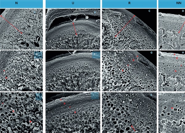
Material and methods: The nourished group received a 20% casein diet, while the undernourished group (U) received a 5% casein diet. The re-nourished groups, R and RR, were used to assess the effects of re-nutrition during puberty and adulthood, respectively. CPs were processed and stained with picro-sirius red, safranin-O and azocarmine. Scanning electron microscopy and immunohistochemistry were also performed.
Results: The area of the CP cartilage and the number of cells in the chondroblastic layer decreased in the U group, as did the thickness of the CP layer in the joint and hypertrophic layer. Renourishment during the pubertal stage, but not during the adult phase, restored these parameters. The cell number was restored when re-nutrition occurred in the pubertal stage, but not in the adult phase. The extracellular matrix also decreased in the U group, but was restored by re-nutrition during the pubertal stage and further increased in the adult phase. IR expression was observed in all CPs, being higher in the chondroblastic and hypertrophic cartilage layers. The lowest expression was found in the U and RR groups.
Conclusions: Protein malnutrition altered the cellularity, the area, and the fibrous cartilage complex, as well as the expression of the IRs.
Figures


Similar articles
-
Bone morphogenetic protein 3 expression pattern in rat condylar cartilage, femoral cartilage and mandibular fracture callus.Eur J Oral Sci. 2005 Aug;113(4):318-25. doi: 10.1111/j.1600-0722.2005.00226.x. Eur J Oral Sci. 2005. PMID: 16048524
-
Parathyroid hormone-related protein regulates proliferation of condylar hypertrophic chondrocytes.J Bone Miner Res. 1999 Nov;14(11):1838-47. doi: 10.1359/jbmr.1999.14.11.1838. J Bone Miner Res. 1999. PMID: 10571683
-
The influence of masseter activity on rat mandibular growth.Arch Oral Biol. 2007 May;52(5):487-93. doi: 10.1016/j.archoralbio.2006.10.019. Epub 2006 Nov 28. Arch Oral Biol. 2007. PMID: 17126288
-
Changes in collagens and chondrocytes in the temporomandibular joint cartilage in growing rats fed a liquid diet.Ann Anat. 2015 Nov;202:78-87. doi: 10.1016/j.aanat.2015.08.006. Epub 2015 Sep 21. Ann Anat. 2015. PMID: 26434755
-
The adaptive remodeling of condylar cartilage---a transition from chondrogenesis to osteogenesis.J Dent Res. 2005 Aug;84(8):691-9. doi: 10.1177/154405910508400802. J Dent Res. 2005. PMID: 16040724 Review.
Cited by
-
Intrauterine growth restriction affects bone mineral density of the mandible and the condyle in growing rats.J Musculoskelet Neuronal Interact. 2022 Mar 1;22(1):93-101. J Musculoskelet Neuronal Interact. 2022. PMID: 35234164 Free PMC article.
-
Age-dependent changes of the mandible bone throughout the lifespan in female F344/N rat.J Anat. 2018 Oct;233(4):440-446. doi: 10.1111/joa.12868. Epub 2018 Aug 2. J Anat. 2018. PMID: 30073652 Free PMC article.
References
-
- Alippi RM, Barcelo AC, Bardi M, Friedman SM, Rio ME, Bozzini C E. Effect of protein-free diet on growth of the skeletal units of the rat mandible. Acta Odontol Latinoam. 1984;1:9–13. - PubMed
-
- Delatte M, Von den Hoff JW, van Rheden RE, Kuijpers-Jagtman AM. Primary and secondary cartilages of the neonatal rat: the femoral head and the mandibular condyle. Eur J Oral Sci. 2004;112:156–162. - PubMed
-
- Fuentes MA, Opperman LA, Bellinger LL, Carlson DS, Hinton RJ. Regulation of cell proliferation in rat mandibular condylar cartilage in explant culture by insulin-like growth factor-1 and fibroblast growth factor-2. Arch Oral Biol. 2002;47:643–654. - PubMed
Publication types
MeSH terms
Substances
LinkOut - more resources
Full Text Sources
Other Literature Sources
Miscellaneous
