. 2015 May 27;11(5):e1004939.
doi: 10.1371/journal.ppat.1004939. eCollection 2015 May.

Selective recruitment of nuclear factors to productively replicating herpes simplex virus genomes

Affiliations

Selective recruitment of nuclear factors to productively replicating herpes simplex virus genomes

Jill A Dembowski et al. PLoS Pathog. .

Abstract

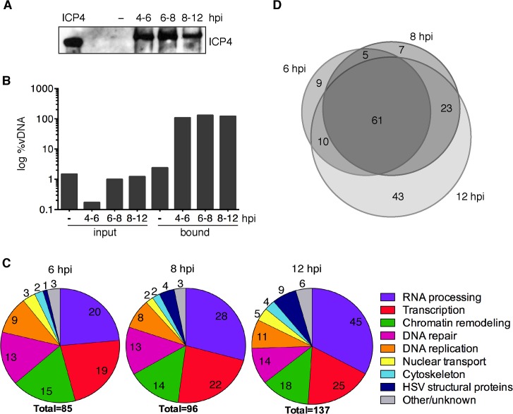
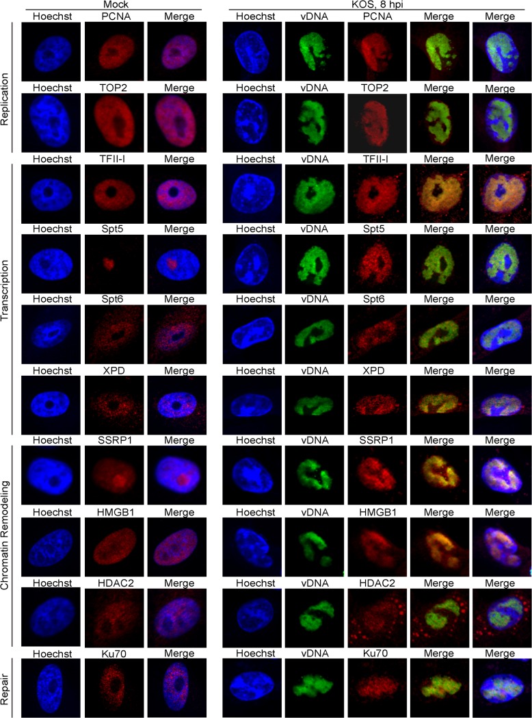
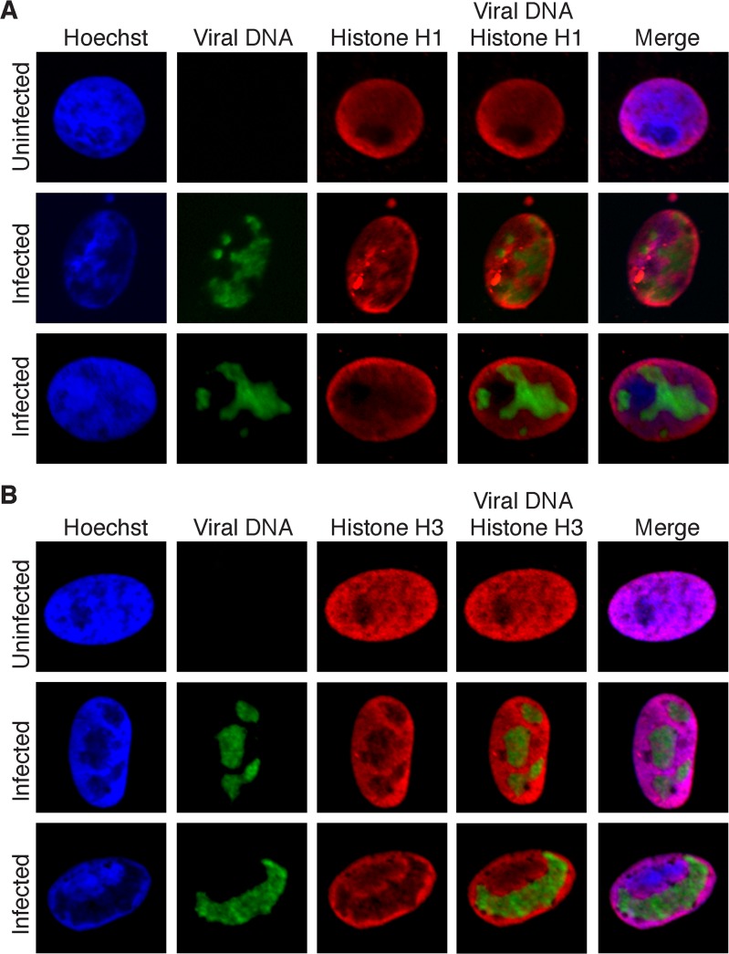
Much of the HSV-1 life cycle is carried out in the cell nucleus, including the expression, replication, repair, and packaging of viral genomes. Viral proteins, as well as cellular factors, play essential roles in these processes. Isolation of proteins on nascent DNA (iPOND) was developed to label and purify cellular replication forks. We adapted aspects of this method to label viral genomes to both image, and purify replicating HSV-1 genomes for the identification of associated proteins. Many viral and cellular factors were enriched on viral genomes, including factors that mediate DNA replication, repair, chromatin remodeling, transcription, and RNA processing. As infection proceeded, packaging and structural components were enriched to a greater extent. Among the more abundant proteins that copurified with genomes were the viral transcription factor ICP4 and the replication protein ICP8. Furthermore, all seven viral replication proteins were enriched on viral genomes, along with cellular PCNA and topoisomerases, while other cellular replication proteins were not detected. The chromatin-remodeling complexes present on viral genomes included the INO80, SWI/SNF, NURD, and FACT complexes, which may prevent chromatinization of the genome. Consistent with this conclusion, histones were not readily recovered with purified viral genomes, and imaging studies revealed an underrepresentation of histones on viral genomes. RNA polymerase II, the mediator complex, TFIID, TFIIH, and several other transcriptional activators and repressors were also affinity purified with viral DNA. The presence of INO80, NURD, SWI/SNF, mediator, TFIID, and TFIIH components is consistent with previous studies in which these complexes copurified with ICP4. Therefore, ICP4 is likely involved in the recruitment of these key cellular chromatin remodeling and transcription factors to viral genomes. Taken together, iPOND is a valuable method for the study of viral genome dynamics during infection and provides a comprehensive view of how HSV-1 selectively utilizes cellular resources.


Conflict of interest statement

The authors have declared that no competing interests exist.

Figures

Fig 1
Fig 1. Schematic representation of procedures used in this paper.
(A) Resting MRC-5 cells in G0 were infected with either prelabeled (left) or unlabeled (right) virus. To assay unreplicated viral DNA (left), prelabeled genomes were processed less than four hpi. To assay viral replication compartments (right), EdU (orange stars) was added to the growth medium during viral DNA replication (≥ 4 hpi) and genomes were assayed 2–4 hours after the addition of EdU. EdU labeled DNA is orange. (B) Viral and cellular DNA, as well as viral and cellular proteins were labeled and visualized as described in the experimental procedures. DNA imaging experiments were carried out in proliferating Vero cells. Viral DNA is green. (C) and (D) iPOND and aniPOND experiments were carried out as described. aniPOND (accelerated native iPOND) is a modified version of iPOND that does not involve crosslinking and therefore requires less stringent wash conditions during purification.
Fig 2
Fig 2. Visualization of EdU labeled HSV-1 genomes.
(A) Prelabeled input viral genomes were visualized in the nucleus of infected cells 2 hpi. Vero cells were infected with wild type KOS or the UL2/UL50 mutant virus carrying unlabeled (0 μM) or prelabeled viral genomes. Labeled virus stocks were generated by growing KOS or UL2/UL50 mutant in the presence of 1.25 or 2.5 μM EdU as described in the experimental procedures. Cellular DNA was visualized by Hoechst staining, viral DNA by click chemistry with EdU, and ICP4 by immunofluorescence. Merged panels show colocalization of viral DNA with ICP4. Prelabeled KOS DNA could not be detected under these conditions. (B) Cells infected with KOS or UL2/UL50 mutant were grown in the presence of 0 or 2.5 μM EdU for 4–8 hpi. Uninfected cells were grown in the presence of EdU for 4 hours. DNA imaging was as described in (A).
Fig 3
Fig 3. iPOND detects viral and cellular proteins associated with replicated HSV-1 genomes.
(A) ICP4 was detected by western blot of protein eluates from iPOND carried out on viral genomes grown in the presence of EdU at 4–6, 6–8, and 8–12 hpi. The control was iPOND carried out on virus grown in the absence of EdU (−) and harvested 8 hpi. Purified ICP4 is shown. (B) DNA eluted from streptavidin-coated beads during iPOND experiments in (A) is viral. The amount of viral DNA present in cell lysates (input) and eluted from beads (bound) during iPOND experiments was measured by qRT-PCR of the viral thymidine kinase (TK) gene. The ratio of viral DNA (vDNA) to total DNA was calculated and is presented as log %vDNA. (C) Pie charts summarize proteins that were identified by mass spectrometry of protein eluates from iPOND carried out 6, 8, and 12 hpi with UL2/UL50 mutant virus. Values indicate the number of proteins identified for each functional category. (D) Venn diagrams depict the overlap of proteins identified by iPOND at each time point.
Fig 4
Fig 4. aniPOND detects viral and cellular proteins associated with replicated viral genomes.
(A) ICP4 was detected in protein eluates from aniPOND experiments carried out on wild type KOS or UL2/UL50 mutant virus by western blotting. AniPOND was carried out on virus grown in the presence (+) or absence (-) of 2.5 μM EdU at 4–8 hpi and/or 33 μM acycloguanosine (ACG) throughout infection. (B) DNA eluted from streptavidin-coated beads during aniPOND experiments in (A) is viral. The amount of viral DNA present in cell lysates (input) and eluted from beads (bound) during aniPOND experiments was measured by qPCR of the viral TK gene. The ratio of viral DNA (vDNA) to total DNA was calculated and is presented in log %vDNA. Values for virus grown in the presence of ACG are too small to be displayed on this graph. These experiments were carried out with the UL2/UL50 mutant virus. (C) Pie charts summarize proteins that were identified by mass spectrometry of protein eluates from aniPOND carried out on the UL2/UL50 mutant grown in the presence of EdU at 4–8 hpi. Pie charts represent proteins that were identified with high confidence in independent duplicate experiments. Values indicate the number of proteins identified for each functional category. (D) Venn diagrams depict the overlap of proteins identified by iPOND and aniPOND carried out on the UL2/UL50 mutant 8 hpi.
Fig 5
Fig 5. Proteins identified to interact with HSV-1 genomes by iPOND and aniPOND relocalize to viral replication compartments during lytic infection.
Vero cells infected with wild type KOS were grown in the presence of 10 μM EdU at 4–8 hpi. Cellular DNA was visualized by Hoechst staining, viral replication compartments (vDNA) by click chemistry with EdU, and cellular proteins by immunofluorescence at 8 hpi (panels KOS, 8 hpi). Mock infected cells (panels Mock) display the normal distribution of cellular proteins in the nucleus in the absence of HSV infection. For KOS infected cells, the first merged panel displays colocalization of viral DNA with cellular proteins. The second merged panel shows the localization of viral DNA and cellular proteins with respect to cellular DNA.
Fig 6
Fig 6. Histones H1 and H3 do not colocalize with replicated HSV genomes.
(A) Infected Vero cells were maintained in the presence of EdU at 4–8 hpi. Cellular DNA was visualized by Hoechst staining, viral replication compartments by click chemistry with EdU, and histone H1 by immunofluorescence. Merged panels show the lack of colocalization of viral DNA with histone H1. Uninfected cells are shown as a control for normal histone distribution in the nucleus. (B) Imaging was carried out as in (A) except that immunofluorescence was carried out with antibodies specific for histone H3.
Fig 7
Fig 7. Histones H1 and H3 do not colocalize with incoming HSV genomes.
Vero cells infected with KOS virus (prelabeled with 10 μM EdU) were assayed for colocalization with histones H1 and H3 at 2 hpi. Nuclei are shown with incoming viral genomes visualized by click chemistry with EdU, and histone H1, histone H3, or ICP4 by immunofluorescence. Merged panels show the lack of colocalization of viral DNA with histones and robust colocalization with ICP4.

References

    1. McGeoch DJ, Dalrymple MA, Davison AJ, Dolan A, Frame MC, et al. (1988) The complete DNA sequence of the long unique region in the genome of herpes simplex virus type 1. J Gen Virol 69: 1531–1574. - PubMed
    1. McGeoch DJ, Dolan A, Donald S, Brauer DH (1986) Complete DNA sequence of the short repeat region in the genome of herpes simplex virus type 1. Nucleic Acids Res 14: 1727–1745. - PMC - PubMed
    1. Batterson W, Furlong D, Roizman B (1983) Molecular genetics of herpes simplex virus. VIII. further characterization of a temperature-sensitive mutant defective in release of viral DNA and in other stages of the viral reproductive cycle. J Virol 45: 397–407. - PMC - PubMed
    1. Ojala PM, Sodeik B, Ebersold MW, Kutay U, Helenius A (2000) Herpes simplex virus type 1 entry into host cells: reconstitution of capsid binding and uncoating at the nuclear pore complex in vitro. Mol Cell Biol 20: 4922–4931. - PMC - PubMed
    1. Shahin V, Hafezi W, Oberleithner H, Ludwig Y, Windoffer B, et al. (2006) The genome of HSV-1 translocates through the nuclear pore as a condensed rod-like structure. J Cell Sci 119: 23–30. - PubMed

Publication types

MeSH terms