. 2015 May;38(5):426-31.
doi: 10.14348/molcells.2015.2288. Epub 2015 May 15.

In Vivo Expression of the PTB-deleted Odin Mutant Results in Hydrocephalus

Affiliations

In Vivo Expression of the PTB-deleted Odin Mutant Results in Hydrocephalus

Sunjung Park et al. Mol Cells. 2015 May.

Abstract

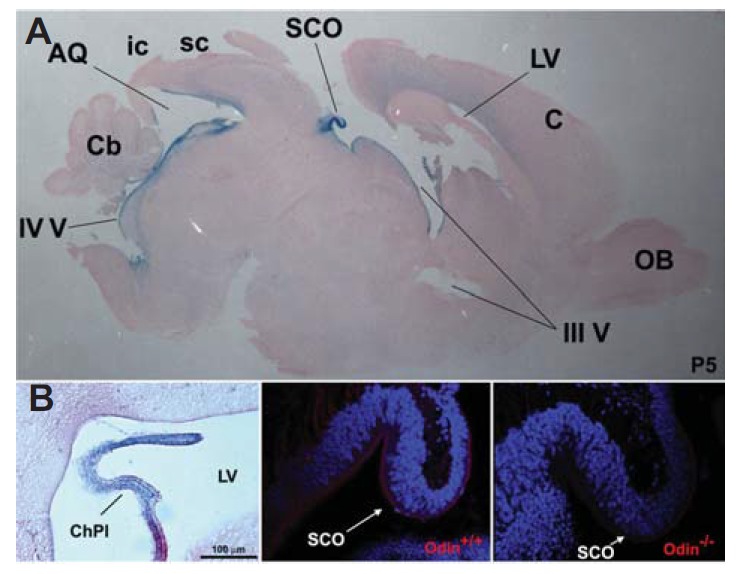
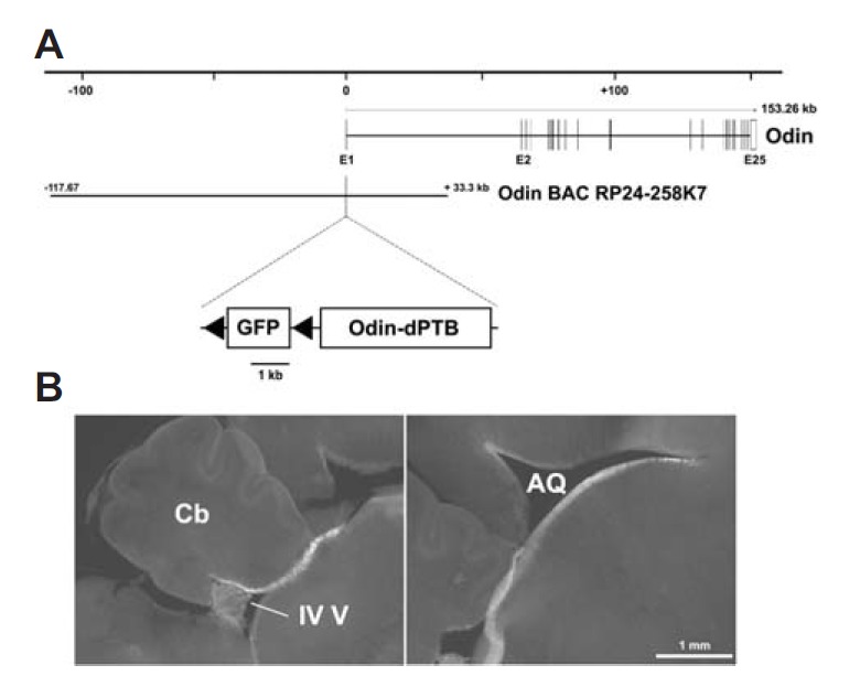
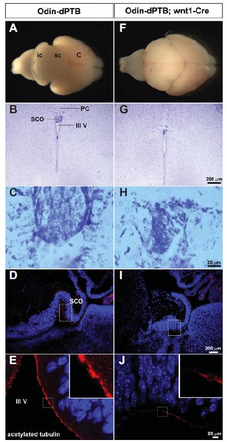
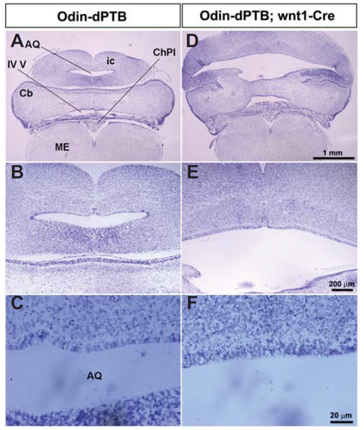
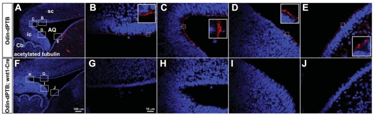
Odin has been implicated in the downstream signaling pathway of receptor tyrosine kinases, such as the epidermal growth factor and Eph receptors. However, the physiologically relevant function of Odin needs to be further determined. In this study, we used Odin heterozygous mice to analyze the Odin expression pattern; the targeted allele contained a β-geo gene trap vector inserted into the 14th intron of the Odin gene. Interestingly, we found that Odin was exclusively expressed in ependymal cells along the brain ventricles. In particular, Odin was highly expressed in the subcommissural organ, a small ependymal glandular tissue. However, we did not observe any morphological abnormalities in the brain ventricles or ependymal cells of Odin null-mutant mice. We also generated BAC transgenic mice that expressed the PTB-deleted Odin (dPTB) after a floxed GFP-STOP cassette was excised by tissue-specific Cre expression. Strikingly, Odin-dPTB expression played a causative role in the development of the hydrocephalic phenotype, primarily in the midbrain. In addition, Odin-dPTB expression disrupted proper development of the subcommissural organ and interfered with ependymal cell maturation in the cerebral aqueduct. Taken together, our findings strongly suggest that Odin plays a role in the differentiation of ependymal cells during early postnatal brain development.

Keywords: Odin; ependymal cells; hydrocephalus; subcommissural organ.


Figures

Fig. 1.
Fig. 1.
Odin expression analysis during early postnatal brain development. (A) The whole brain was dissected out of Odin heterozygous mice at postnatal (P) day 5 and processed for cryostat sections. The mid-sagittal section was subjected to X-gal staining to reveal LacZ-positive cells lining the brain ventricle. AQ, cerebral aqueduct; C, cerebral cortex; Cb, cerebellum; ic, inferior colliculus; LV, lateral ventricle; OB, olfactory bulb; sc, superior colliculus; SCO, subcommissural organ; III V, third ventricle; IV V, fourth ventricle. (B) LacZ expression analysis of the choroid plexus (ChPl) found in the lateral ventricle (LV). (C and D) The mid-sagittal section from the indicated Odin mice was subjected to immunohistochemical staining using anti-Odin antibodies.
Fig. 2.
Fig. 2.
Green fluorescent protein (GFP) expression analysis using Odin BAC transgenic mice. (A) Schematic map of the mouse Odin genomic locus with the Odin BAC clone (RP24-258K7). The recombinant Odin BAC clone contained a floxed GFP-STP cassette followed by the Odin-dPTB expression cassette inserted upstream into the start codon of the first exon. (B) GFP fluorescent image of the mid-sagittal section prepared by vibratome (100 μm-thick). Similar results were reproducibly observed in three different transgenic mice.
Fig. 3.
Fig. 3.
Subcommissural organ (SCO) development is defective in mice ectopically expressing Odin-dPTB. Littermate mice at P5 were obtained from a cross between the Odin-dPTB BAC transgenic line and wnt1-Cre mice to analyze their morphological abnormalities. (A and F) Whole brains were dissected out of the indicated mice for morphological comparison. Note that the brain of the double transgenic mice reveals a severe hydrocephalic phenotype with prominent enlargement in the midbrain. (B and G) Cresyl violet staining analysis of the coronal section. Note that the SCO is much smaller in the brain of the double transgenic line. PC, posterior commissure. (C and H) Enlarged view of the SCO regions in B and G, respectively. (D and I) The mid-sagittal section was subjected to immunohistochemical staining using anti-acetylated tubulin antibody, a marker for multicilia of ependymal cells. (E and J) Enlarged view of the SCO regions in D and I, respectively. Note that the ependymal cells in the SCO display much weaker staining with anti-acetylated tubulin antibody.
Fig. 4.
Fig. 4.
The midbrain of Odin-dPTB mice becomes severely expanded upon Cre expression. (A and D) The coronal sections of the indicated brains were analyzed by cresyl violet staining. Note that both the cerebral aqueduct and inferior colliculus (ic) are severely expanded. ME, medulla. (B and E) Enlarged view of the cerebral aqueduct regions in A and D, respectively. (C and F) High magnification of the ependymal cells lining the cerebral aqueduct in B and E. Note that the ependymal cells of control mice have a regular and well organized morphology, whereas those of the double transgenic mice do not.
Fig. 5.
Fig. 5.
The ependymal cells lining the cerebral aqueduct are defective for development of multi-cilia. (A and F) The mid-sagittal section of the indicated mice was subjected to immunohistochemical staining using anti-acetylated antibody to reveal multi-ciliated ependymal cells. (B–E and G–J) High magnification of the brain regions corresponding to boxes in A and F, respectively.

References

    1. Baas D, Meiniel A, Benadiba C, Bonnafe E, Meiniel O, Reith W, Durand B. A deficiency in RFX3 causes hydrocephalus associated with abnormal differentiation of ependymal cells. Eur. J. Neurosci. 2006;24:1020–1030. - PubMed
    1. Blatt EN, Yan XH, Wuerffel MK, Hamilos DL, Brody SL. Forkhead transcription factor HFH-4 expression is temporally related to ciliogenesis. Am. J. Respir. Cell Mol. Biol. 1999;21:168–176. - PubMed
    1. Breunig JJ, Arellano JI, Rakic P. Cilia in the brain: going with the flow. Nat. Neurosci. 2010;13:654–655. - PubMed
    1. Carlen M, Meletis K, Goritz C, Darsalia V, Evergren E, Tanigaki K, Amendola M, Barnabe-Heider F, Yeung MS, Naldini L, et al. Forebrain ependymal cells are Notch-dependent and generate neuroblasts and astrocytes after stroke. Nat. Neurosci. 2009;12:259–267. - PubMed
    1. Cottrell GT, Ferguson AV. Sensory circumventricular organs: central roles in integrated autonomic regulation. Regul. Pept. 2004;117:11–23. - PubMed

Publication types

MeSH terms

Substances

LinkOut - more resources