. 2015 May 28:6:7205.
doi: 10.1038/ncomms8205.

Abnormal splicing switch of DMD's penultimate exon compromises muscle fibre maintenance in myotonic dystrophy

Affiliations

Abnormal splicing switch of DMD's penultimate exon compromises muscle fibre maintenance in myotonic dystrophy

Frédérique Rau et al. Nat Commun. .

Abstract

Myotonic Dystrophy type 1 (DM1) is a dominant neuromuscular disease caused by nuclear-retained RNAs containing expanded CUG repeats. These toxic RNAs alter the activities of RNA splicing factors resulting in alternative splicing misregulation and muscular dysfunction. Here we show that the abnormal splicing of DMD exon 78 found in dystrophic muscles of DM1 patients is due to the functional loss of MBNL1 and leads to the re-expression of an embryonic dystrophin in place of the adult isoform. Forced expression of embryonic dystrophin in zebrafish using an exon-skipping approach severely impairs the mobility and muscle architecture. Moreover, reproducing Dmd exon 78 missplicing switch in mice induces muscle fibre remodelling and ultrastructural abnormalities including ringed fibres, sarcoplasmic masses or Z-band disorganization, which are characteristic features of dystrophic DM1 skeletal muscles. Thus, we propose that splicing misregulation of DMD exon 78 compromises muscle fibre maintenance and contributes to the progressive dystrophic process in DM1.


Figures
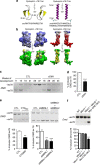
Figure 1
Figure 1. DMD exon 78 MBNL-regulated splicing switch changes dystrophin C-ter tail.
(a) PEP-fold analysis of dystrophin +78 and dystrophin Δ78 C-ter structures. (b) Surface properties of PEP-fold models of dystrophin +78 and dystrophin Δ78 C-ter. Electrostatic potentials (upper panel) are shown in blue for electropositive and red for electronegative. Hydrophobic and hydrophilic surface potentials (lower panel) are coloured in yellow and green, respectively. (c) RT–PCR analysis of DMD exon 78 alternative splicing in human skeletal muscle samples from control (CTL) and congenital DM1 (cDM1) fetuses. (d) Quantification of DMD exon 78 inclusion in skeletal muscle samples from cDM1 compared with control fetuses aged between 20 and 37 weeks of development (n=3). (e) Upper left panel: RT–PCR analysis of endogenous DMD exon 78 inclusion in control differentiated human muscle cells with or without the expression of conditional 960 CTG repeats. Upper right panel: RT–PCR analysis of endogenous DMD exon 78 mRNA in control differentiated human muscle cells transfected with siRNAs against MBNL1 or both MBNL1 and MBNL2. Lower panel: quantification of DMD exon 78 inclusion (n=6 from three independent experiments). (f) RT–PCR analysis and quantification of endogenous Dmd mRNA in skeletal muscle samples from WT, Mbnl1−/− Knockout, Mbnl1−/−, Mbnl2+/− Knockout and myo-CRE muscle-specific Mbnl1−/−, Mbnl2−/− double Knockout (myo-CRE DKO) (n=3). Bars indicates s.e.m. and ** indicates P<0.01; *** indicates P<0.001; Student's t-test).
Figure 2
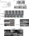
Figure 2. Exclusion of dmd exon 78 in zebrafish impairs skeletal muscle development.
(a) RT–PCR of dmd exon 78 performed on total RNA extracts isolated from whole control and dmd Δ78 embryos (48 hpf). (b) Dose-dependant phenotype of dmd Δ78 embryos: control embryos (CTL) compared with moderate and severe affected dmd Δ78 morphants at 48 hpf (scale bar, 1 mm). (c) Touch-evoked escape test of control embryos compared with moderate and severe dmd Δ78 morphants (1 image/0.2 s; scale bar, 1 mm). (d) Abnormal myoseptum U-shape in dmd Δ78 morphants compared with V-shape in control embryos at 48 hpf (scale bar, 250 and 100 μm). (e) Dystrophin immunostaining (MANDRA1 antibody) of control embryos compared with dmd Δ78 morphants at 48 hpf. (f) Slow myosin immunostaining of control embryos compared with moderate and severe affected dmd Δ78 morphants at 48 hpf (scale bar, 50 and 10 μm).
Figure 3
Figure 3. μ-dystrophin with a 31aa C-ter tail fails to ameliorate mdx muscle phenotype.
(a) Dystrophin N-terminal domain (Manex1011B antibody), dystrophin C-terminus domain (Dys2 antibody), α-sarcoglycan, α-syntrophin and α-dystrobrevin immunostaining of mdx4cv TA muscles injected with saline, AAV2/9-μDys-CTL or AAV2/9-μDys-Δex78 (scale bar, 50 μm). (b,e) TA muscle weight of C57BL/6 control mice (C57, n=8) compared with TA muscles of mdx4cv mice (n=8) injected with saline (mdx4cv) or AAV2/9-μDys-CTL (b) or AAV2/9-μDys-Δex78 (e). (c,f) Specific maximal force (sP0) of TA muscles of C57BL/6 control mice (C57, n=8) compared with TA muscles of mdx4cv mice (n=8) injected with saline (mdx4cv) or AAV2/9-μDys-CTL (c) or AAV2/9-μDys-Δex78 (f). (d,g) Resistance to eccentric contractions. Absolute maximal force (P0) following lengthening contractions of TA muscles of C57BL/6 control mice (C57, n=8) compared with TA muscles of mdx4cv mice (n=8) injected with saline (mdx4cv) or AAV2/9-μDys-CTL (d) or AAV2/9-μDys-Δex78 (g). Bars indicate s.e.m. and ‘NS' indicates not significant; * indicates P<0.05; ** indicates P<0.01 compared to mdx4cv condition; one-way analysis of variance with Tukey's multiple comparisons test.
Figure 4
Figure 4. Dystrophin exon 78 is required for muscle structure maintenance.
(a) RT–PCR analysis and quantification of Dmd exon 78 inclusion in TA muscles (n=10) injected with AAV-U7-Dmd-ex78 compared with contralateral TA muscles injected with saline (CTL). (b) Hematoxylin and eosin staining in TA muscles injected either with saline (CTL) or AAV-U7-Dmd-ex78 (scale bar, 50 μm). Inset: higher magnification (× 2.5) of a structure reminiscent of ringed fibre shown by the arrow. (c) Fibre types in TA muscles injected either with saline (CTL) or AAV-U7-Dmd-ex78 were determined by MyHC immunostaining: MyHC-IIa in red, MyHC-IIx in blue, MyHC-IIb dark, laminin in green. Purple fibres correspond to MyHC-IIa and MyHC-IIx positive fibres (scale bar, 100 μm). (d) Quantification of MyHC mRNA levels by quantitative RT–PCR (n=10). Bars indicate s.e.m. and ‘NS' indicates not significant; *** indicates P<0.001; Student's t-test). (e) Ultrastructure of representative, longitudinally cut, fibres in AAV-U7-Dmd-ex78-injected TA. Upper panel: ‘ringed fibre'. Sarcomeres are mainly longitudinally oriented, but just under the sarcolemma, a band of myofibrils (pseudo-coloured in green) is transversally oriented as evidenced in the enlarged zone (N, nucleus; M, mitochondria). Lower panel: sarcoplasmic mass. The sarcoplasm beneath its sarcolemma appears nearly devoid of myofilaments and the higher magnification shows some electrodense remnants of Z line material and vacuoles of swollen sarcoplasmic reticulum. In addition focal zones with Z line streaming are also observed in fibres of AAV-U7-Dmd-ex78-injected muscles (arrows in left panels). Left panels scale bars, 5 μm and rigth panels scale bars, 1 μm.

References

    1. Brook J. D. et al.. Molecular basis of myotonic dystrophy: expansion of a trinucleotide (CTG) repeat at the 3' end of a transcript encoding a protein kinase family member. Cell 69, 385 (1992). - PubMed
    1. Fu Y. H. et al.. An unstable triplet repeat in a gene related to myotonic muscular dystrophy. Science 255, 1256–1258 (1992). - PubMed
    1. Mahadevan M. et al.. Myotonic dystrophy mutation: an unstable CTG repeat in the 3' untranslated region of the gene. Science 255, 1253–1255 (1992). - PubMed
    1. Kanadia R. N. et al.. A muscleblind knockout model for myotonic dystrophy. Science 302, 1978–1980 (2003). - PubMed
    1. Kanadia R. N. et al.. Developmental expression of mouse muscleblind genes Mbnl1, Mbnl2 and Mbnl3. Gene Expr. Patterns 3, 459–462 (2003). - PubMed

Publication types

MeSH terms