. 2015 May 27;10(5):e0126589.
doi: 10.1371/journal.pone.0126589. eCollection 2015.

Lethal interpersonal violence in the Middle Pleistocene

Affiliations

Lethal interpersonal violence in the Middle Pleistocene

Nohemi Sala et al. PLoS One. .

Abstract

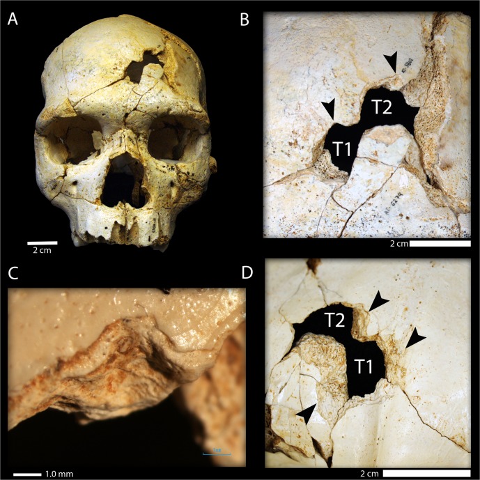
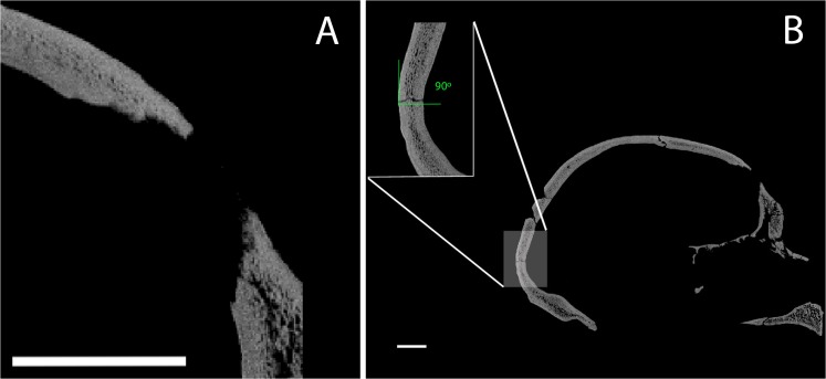
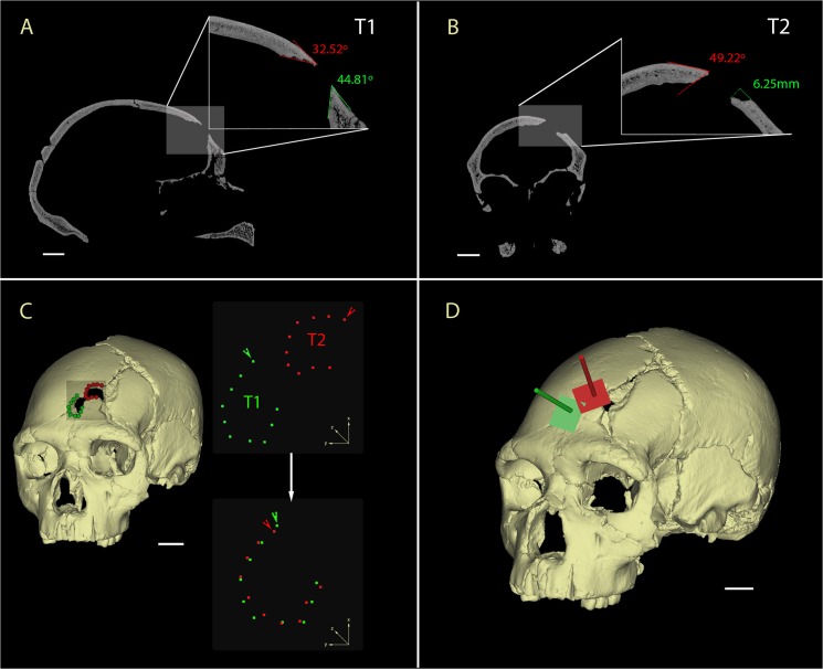
Evidence of interpersonal violence has been documented previously in Pleistocene members of the genus Homo, but only very rarely has this been posited as the possible manner of death. Here we report the earliest evidence of lethal interpersonal violence in the hominin fossil record. Cranium 17 recovered from the Sima de los Huesos Middle Pleistocene site shows two clear perimortem depression fractures on the frontal bone, interpreted as being produced by two episodes of localized blunt force trauma. The type of injuries, their location, the strong similarity of the fractures in shape and size, and the different orientations and implied trajectories of the two fractures suggest they were produced with the same object in face-to-face interpersonal conflict. Given that either of the two traumatic events was likely lethal, the presence of multiple blows implies an intention to kill. This finding shows that the lethal interpersonal violence is an ancient human behavior and has important implications for the accumulation of bodies at the site, supporting an anthropic origin.


Conflict of interest statement

Competing Interests: The authors have declared that no competing interests exist.

Figures

Fig 1
Fig 1. Stratigraphy of the Sima de los Huesos site (modified from Arsuaga et al. [21]).
The hominin bones were recovered in Lithostratigraphic Unit 6 (LU-6) dated to c. 430ka [21]. This unit is composed of pure red clays, filtering into the conduit system from overlying soils with little or no lateral transport, and very low velocity of sedimentation (decantation by dripping water) [23]. The figure also shows a detailed image of Cr-17 during its excavation at the site. Note the pure red clay that covers the cranial bones (partially cleaned in situ to enhance visualization) and the typical in situ postmortem (fossil diagenetic) fractures of the cranial vault. Photo credit: Javier Trueba (Madrid Scientific Films).
Fig 2
Fig 2. Cranium 17 bone traumatic fractures.
(A) Frontal view of Cranium 17 showing the position of the traumatic events T1 (inferior) and T2 (superior); (B) Detailed ectocranial view of the traumatic fractures showing the two similar notches (black arrows) present along the superior border of the fracture outlines. Note that the orientation of the two traumatic events is different; (C) Detail of the notch in T1 under 2X magnification with a light microscope. (D) Endocranial view of T1 and T2 showing the large cortical delamination of the inner table (black arrows).
Fig 3
Fig 3. CT cross-sections comparing perimortem and postmortem fractures in the SH crania.
(A) Perimortem fracture of the frontal bone in SH Cranium 17; (B) Postmortem fracture in the occipital bone of SH Cranium 17 showing a right angle at the point of breakage. Scale bar = 2 cm.
Fig 4
Fig 4. CT analysis of the Cranium 17 traumas.
A) Sagittal cross-section of the T1 showing the acute fracture angles (32.5°-44.8°); B) Coronal cross-section of the T2 in detail showing the acute fracture angle (49.2°) and the large cortical delamination of the outer table (6.25mm); C) Virtual reconstruction of the SH Cranium 17 (left) showing the fractures on the left frontal squama and the landmarks used for the shape analysis (blue = T1 and red = T2). 3D landmarks of T1 and T2 in their original position (upper right), with arrows indicating the first landmark placed on the notch. Rotation and superposition of the 3D landmarks of T1 and T2 using the first landmark of the two fractures as reference points (lower right). D) Reconstruction of the trajectory of the impacts relying on the planar orientations of the external outlines of the fractures (blue = T1 and red = T2). Scale bars 2 cm.

References

    1. Martin DL, Harrod RP. Bioarchaeological contributions to the study of violence. Yearb Phys Anthropol. 2015; 156: 116–145. - PubMed
    1. Walker PL. Cranial injuries as evidence of violence in prehistoric southern California. Am J Phys Anthropol. 1989; 80: 313–323. - PubMed
    1. Jiménez-Brobeil SA, Oumaoui A. Possible relationship of cranial traumatic injuries with violence in the South-East Iberian Peninsula from the Neolithic to the Bronze Age. Am J Phys Anthropol. 2009; 140. - PubMed
    1. Smith MO. Beyond palisades: The nature and frequency of late prehistoric deliberate violent trauma in the Chickamauga Reservoir of East Tennessee. Am J Phys Anthropol. 2003; 121: 303–318. - PubMed
    1. Spencer SD. Detecting violence in the archaeological record: Clarifying the timing of trauma and manner of death in cases of cranial blunt force trauma among pre-Columbian Amerindians of West-Central Illinois. Int J Paleopathol. 2012; 2: 112–122. - PubMed

Publication types

LinkOut - more resources