Multiscale mechanobiology: computational models for integrating molecules to multicellular systems
- PMID: 26019013
- PMCID: PMC4677065
- DOI: 10.1039/c5ib00043b
Multiscale mechanobiology: computational models for integrating molecules to multicellular systems
Abstract
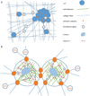
Mechanical signals exist throughout the biological landscape. Across all scales, these signals, in the form of force, stiffness, and deformations, are generated and processed, resulting in an active mechanobiological circuit that controls many fundamental aspects of life, from protein unfolding and cytoskeletal remodeling to collective cell motions. The multiple scales and complex feedback involved present a challenge for fully understanding the nature of this circuit, particularly in development and disease in which it has been implicated. Computational models that accurately predict and are based on experimental data enable a means to integrate basic principles and explore fine details of mechanosensing and mechanotransduction in and across all levels of biological systems. Here we review recent advances in these models along with supporting and emerging experimental findings.
Figures


Similar articles
-
Brushes, cables, and anchors: recent insights into multiscale assembly and mechanics of cellular structural networks.Cell Biochem Biophys. 2007;47(3):348-60. doi: 10.1007/s12013-007-0013-x. Cell Biochem Biophys. 2007. PMID: 17652780 Review.
-
Force localization modes in dynamic epithelial colonies.Mol Biol Cell. 2018 Nov 15;29(23):2835-2847. doi: 10.1091/mbc.E18-05-0336. Epub 2018 Sep 12. Mol Biol Cell. 2018. PMID: 30207837 Free PMC article.
-
Single cell active force generation under dynamic loading - Part II: Active modelling insights.Acta Biomater. 2015 Nov;27:251-263. doi: 10.1016/j.actbio.2015.09.004. Epub 2015 Sep 7. Acta Biomater. 2015. PMID: 26360595
-
Mechanobiology of collective cell behaviours.Nat Rev Mol Cell Biol. 2017 Dec;18(12):743-757. doi: 10.1038/nrm.2017.98. Epub 2017 Nov 8. Nat Rev Mol Cell Biol. 2017. PMID: 29115298 Review.
-
United we stand: integrating the actin cytoskeleton and cell-matrix adhesions in cellular mechanotransduction.J Cell Sci. 2012 Jul 1;125(Pt 13):3051-60. doi: 10.1242/jcs.093716. Epub 2012 Jul 13. J Cell Sci. 2012. PMID: 22797913 Free PMC article. Review.
Cited by
-
Engineering innovations in medicine and biology: Revolutionizing patient care through mechanical solutions.Heliyon. 2024 Feb 15;10(4):e26154. doi: 10.1016/j.heliyon.2024.e26154. eCollection 2024 Feb 29. Heliyon. 2024. PMID: 38390063 Free PMC article. Review.
-
A stochastic algorithm for accurately predicting path persistence of cells migrating in 3D matrix environments.PLoS One. 2018 Nov 15;13(11):e0207216. doi: 10.1371/journal.pone.0207216. eCollection 2018. PLoS One. 2018. PMID: 30440015 Free PMC article.
-
Risky interpretations across the length scales: continuum vs. discrete models for soft tissue mechanobiology.Biomech Model Mechanobiol. 2022 Apr;21(2):433-454. doi: 10.1007/s10237-021-01543-4. Epub 2022 Jan 5. Biomech Model Mechanobiol. 2022. PMID: 34985590 Free PMC article.
-
A constriction channel analysis of astrocytoma stiffness and disease progression.Biomicrofluidics. 2021 Mar 16;15(2):024103. doi: 10.1063/5.0040283. eCollection 2021 Mar. Biomicrofluidics. 2021. PMID: 33763160 Free PMC article.
-
Mechanobiology of the female reproductive system.Reprod Med Biol. 2021 Jul 31;20(4):371-401. doi: 10.1002/rmb2.12404. eCollection 2021 Oct. Reprod Med Biol. 2021. PMID: 34646066 Free PMC article. Review.
References
-
- Szabó B, Szöllösi GJ, Gönci B, Jurányi Z, Selmeczi D, Vicsek T. Phase transition in the collective migration of tissue cells: experiment and model, Phys. Rev. E: Stat., Nonlinear. Soft Matter Phys. 2006;74:061908. - PubMed
Publication types
MeSH terms
Substances
Grants and funding
LinkOut - more resources
Full Text Sources
Other Literature Sources

