. 2015 May 27;35(21):8359-75.
doi: 10.1523/JNEUROSCI.2850-14.2015.

Exclusion of integrins from CNS axons is regulated by Arf6 activation and the AIS

Affiliations

Exclusion of integrins from CNS axons is regulated by Arf6 activation and the AIS

Elske H P Franssen et al. J Neurosci. .

Abstract

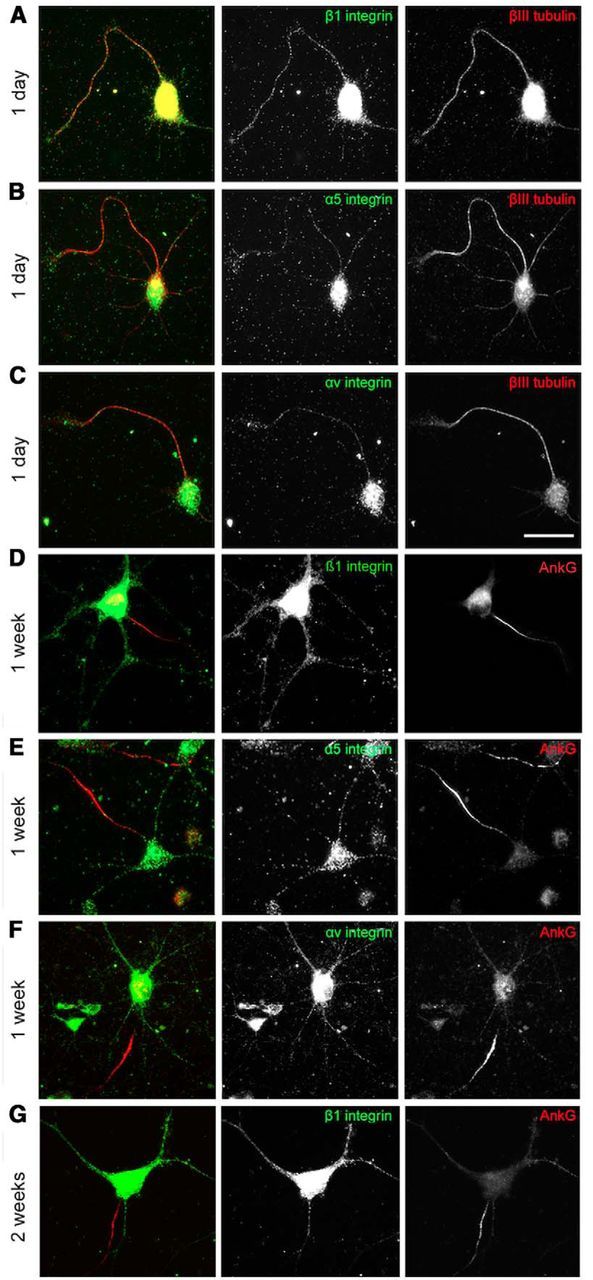
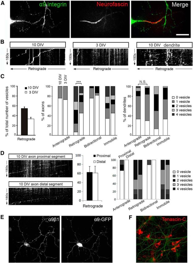
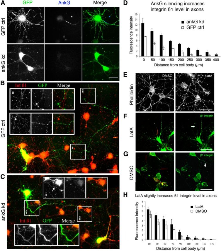
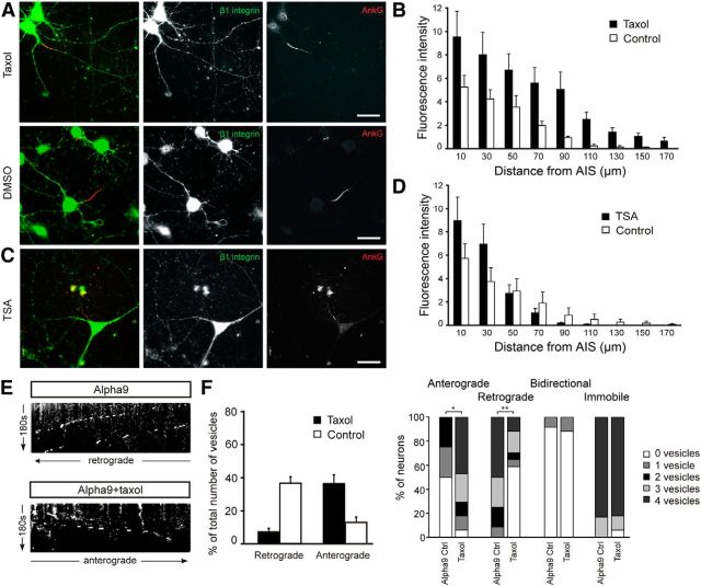
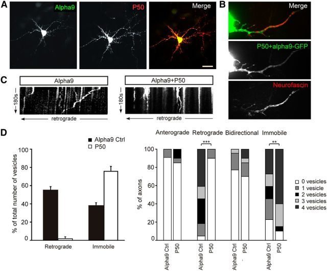
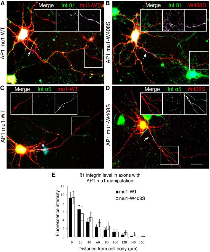
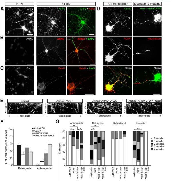
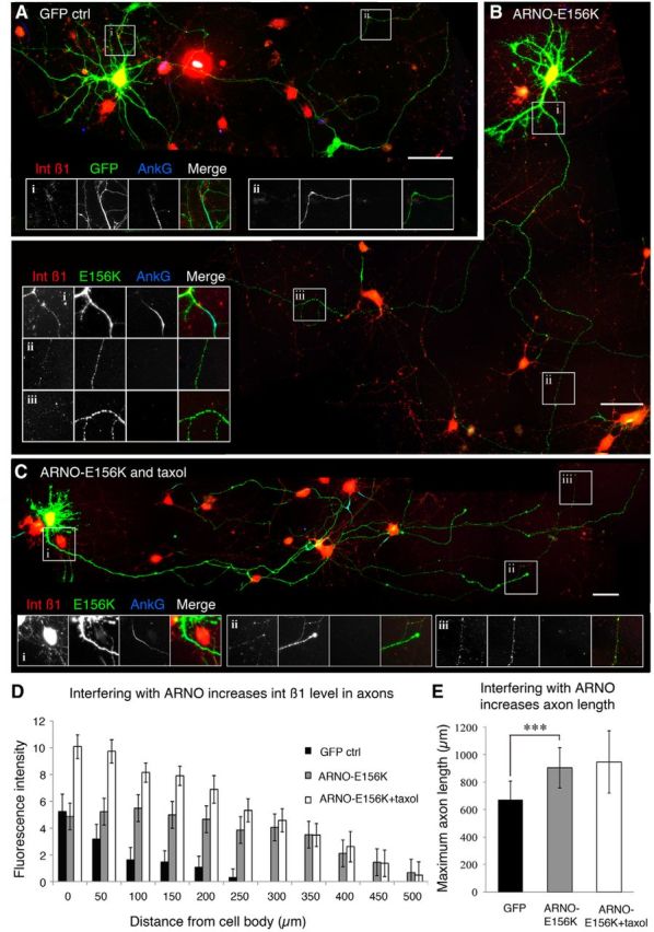
Integrins are adhesion and survival molecules involved in axon growth during CNS development, as well as axon regeneration after injury in the peripheral nervous system (PNS). Adult CNS axons do not regenerate after injury, partly due to a low intrinsic growth capacity. We have previously studied the role of integrins in axon growth in PNS axons; in the present study, we investigate whether integrin mechanisms involved in PNS regeneration may be altered or lacking from mature CNS axons by studying maturing CNS neurons in vitro. In rat cortical neurons, we find that integrins are present in axons during initial growth but later become restricted to the somato-dendritic domain. We investigated how this occurs and whether it can be altered to enhance axonal growth potential. We find a developmental change in integrin trafficking; transport becomes predominantly retrograde throughout axons, but not dendrites, as neurons mature. The directionality of transport is controlled through the activation state of ARF6, with developmental upregulation of the ARF6 GEF ARNO enhancing retrograde transport. Lowering ARF6 activity in mature neurons restores anterograde integrin flow, allows transport into axons, and increases axon growth. In addition, we found that the axon initial segment is partly responsible for exclusion of integrins and removal of this structure allows integrins into axons. Changing posttranslational modifications of tubulin with taxol also allows integrins into the proximal axon. The experiments suggest that the developmental loss of regenerative ability in CNS axons is due to exclusion of growth-related molecules due to changes in trafficking.

Keywords: Arf6; axon initial segment; axonal transport; integrin; trafficking.


Figures

Figure 1.
Figure 1.
Integrins are not transported into the axons of mature CNS neurons. AC, At 1 DIV, various types of integrins (β1, α5, and αv) are distributed ubiquitously in cell bodies and neurites. D, At 7 DIV, the β1 integrin level is gradually reduced in axons and dendrites. At 1 week (E, F), α integrins are restricted to the somatodendritic domain and absent in axons. At 2 weeks (G), β1 integrins disappear from axons too. Scale bars: AC, 25 μm; DG, 50 μm.
Figure 2.
Figure 2.
In mature CNS axons, α9-GFP vesicles are retrogradely transported to the cell body. A, Day 10 cortical neuron transfected with α9-GFP was stained with Neufascin to identify the axon, where the vesicle movement was imaged. Transfected α9-GFPs are slightly missorted, with more integrin molecules found in the axons. Scale bar, 25 μm. B, Kymographs of α9-GFP vesicles traffic in axons at 10 DIV (left) and 3 DIV (middle) and in a dendrite at 10 DIV (right). In axons, less retrograde transportation was observed in younger cultures compared with 10 DIV cultures; in dendrites, vesicles were transported in both directions at 10 DIV. C, From 3 to 10 DIV (left), the overall percentage of retrogradely moving vesicles increases; a greater percentage of axons were observed to contain retrogradely moving vesicles and the number of vesicles increased as axons mature (middle; ***p < 0.001, Mann–Whitney test); in dendrites at 10 DIV, no significant difference was observed between number of anterogradely and retrogradely moving α9 vesicles (right). D, Kymographs and analysis of α9-GFP vesicle traffic in axons at 10 DIV at a region 100 μm into the axon (proximal) and toward the end of the axon (distal, 300–600 μm). E, α9β1 dimer was formed in neurons transfected with α9-GFP. F, Neuron cultures produce tenascin-C on the substrate. Scale bars, 50 μm.
Figure 3.
Figure 3.
Disruption of the AIS increased β1 integrin expression in axons. A, Adenovirus-mediated delivery of AnkG shRNA resulted in silencing of AnkG in cortical neurons 72 h after infection. BD, At 72 h after transduction, integrin β1 level is increased in axon (C) compared with GFP control (B) by 3.917 ± 0.369 units (p < 0.001, group difference by Tukey post hoc; D). E, LatA results in disruption of actin filaments (phalloidin staining) and results in a small but significant increase in β1 integrin level in the axons (FH; p = 0.002). Note that the experimental proceddures of LatA treatment and AnkG knock down were very different, the measurements are not to be compared between experiments. Scale bars, A-C, 25 μm. EG, 50 μm.
Figure 4.
Figure 4.
Tubulin PTM modification results in increased axonal β1 integrin. A, B, Low-dose taxol treatment increased β1 integrin level in the axons with group difference of 3.149 ± 0.697 arbitrary units (p < 0.001). C, D, TSA-induced increased acetylation did not increase β1 integrin level in the axons. E, F, Taxol treatment increased α9-GFP vesicle anterograde movement. **p < 0.01; *p < 0.05. Scale bars in AC, 25 μm.
Figure 5.
Figure 5.
Retrograde transport of α9-GFP in axons is mediated by dyneins. A, Neurons were cotransfected with α9-GFP and p50-HA. Scale bar, 30 μm. B, Axons were identified with Neurofascin live staining. C, D, Inhibiting dynein function resulted in an abolishment of retrograde transport but increased immobile vesicles ***p < 0.001; **p < 0.01.
Figure 6.
Figure 6.
AP-1-dependent somatodendritic sorting is not the main mechanism for integrin axonal exclusion. Cortical neurons were transfected with WT (A, C) and the mutated (B, D) AP-1 mu1 subunit and the integrin level was measured in axons with β1 (A, B) and α5 (C, D). Arrows point to the AIS, which is stained with AnkG in blue and is used to identify axon branches. E, Expression of mutated AP-1 did not increase the concentration of β1 or a5 integrins in axons and did not allow integrin α5 to enter the axon far beyond axon initial segments. Scale bars, 25 μm.
Figure 7.
Figure 7.
Direction of axonal integrin transport in mature CNS axons is controlled by ARF6. A, ARF6 was present in axons and dendrites of neurons at both 2 and 14 DIV. B, ARNO is increased in axons and dendrites of 14DIV neurons compared with 2 DIV and is expressed throughout the axon (MAP2 negative). C, Rab11 is present in dendrites but not in axons of 14 DIV neurons. D, For live imaging, neurons were cotransfected with α9-GFP and either ACAP1-myc or ARNO-E156K-myc and axons were identified with neurofascin. E, F, G, Kymographs and analysis of α9-GFP vesicle traffic in control axons or after transfection with ACAP1, ARNO-E156K, or ARNO-E156K combined with taxol treatment. ***p < 0.001; **p < 0.01; *p < 0.05.
Figure 8.
Figure 8.
ARNO-E156K significantly increased integrin level in axons as well as axon length. A, In day 9 cortical neurons transfected with GFP, integrin β1 was absent in axons beyond the AIS (stained with AnkG). B, Overexpression of ARNO-E156K increased β1 integrin level in axons including in the distal part (ii,iii). C, Combination of ARNO-E156K with taxol treatment showed a similar effect. D, Integrin β1 fluorescent intensity in axons is increased with ARNO-E156K or the combination treatment (ARNO-E156K vs GFP: 3.168 ± 0.362; ARNO-E156K + taxol vs GFP: 6.223 ± 1.436; p < 0.001 in both cases). Compared with ARNO-E156K alone, the combination increased integrin β1 in the proximal 200 μm (t = 0.0027, paired t test), but not in the distal part. E, ARNO-E156K significantly increased axon length; combination with taxol had no additional effect. ***p < 0.001. Scale bars in AC, 50 μm.

References

    1. Andrews MR, Czvitkovich S, Dassie E, Vogelaar CF, Faissner A, Blits B, Gage FH, ffrench-Constant C, Fawcett JW. Alpha9 integrin promotes neurite outgrowth on tenascin-C and enhances sensory axon regeneration. J Neurosci. 2009;29:5546–5557. doi: 10.1523/JNEUROSCI.0759-09.2009. - DOI - PMC - PubMed
    1. Arjonen A, Kaukonen R, Ivaska J. Filopodia and adhesion in cancer cell motility. Cell Adh Migr. 2011;5:421–430. doi: 10.4161/cam.5.5.17723. - DOI - PMC - PubMed
    1. Arnold DB. Actin and microtubule-based cytoskeletal cues direct polarized targeting of proteins in neurons. Sci Signal. 2009;2:pe49. - PMC - PubMed
    1. Bi X, Lynch G, Zhou J, Gall CM. Polarized distribution of integrin alpha5 in dendrites of hippocampal and cortical neurons. J Comp Neurol. 2001;435:184–193. doi: 10.1002/cne.1201. - DOI - PubMed
    1. Blackmore MG, Wang Z, Lerch JK, Motti D, Zhang YP, Shields CB, Lee JK, Goldberg JL, Lemmon VP, Bixby JL. Kruppel-like Factor 7 engineered for transcriptional activation promotes axon regeneration in the adult corticospinal tract. Proc Natl Acad Sci U S A. 2012;109:7517–7522. doi: 10.1073/pnas.1120684109. - DOI - PMC - PubMed

Publication types

LinkOut - more resources