Review
. 2015 May 21;21(19):6088-96.
doi: 10.3748/wjg.v21.i19.6088.

Primary hepatic angiosarcoma: A report of two cases and literature review

Affiliations
Review

Primary hepatic angiosarcoma: A report of two cases and literature review

Yi-Ping Zhu et al. World J Gastroenterol. .

Abstract

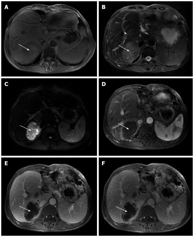
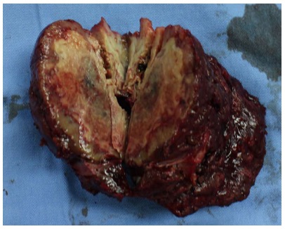
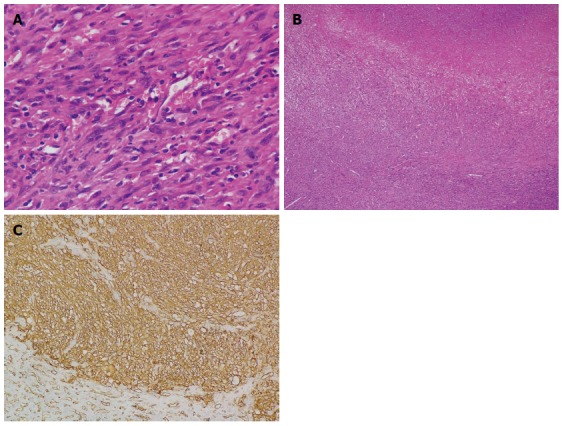
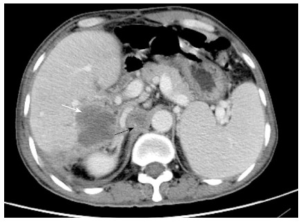
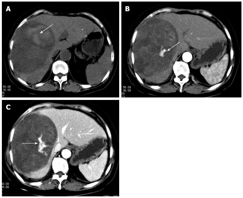
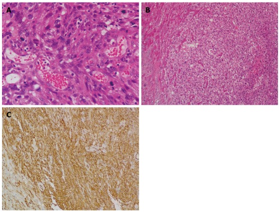
Primary hepatic angiosarcoma (PHA) is a rare malignancy that carries a poor prognosis. Of 1500 patients who underwent hepatectomy for primary hepatic tumors between 1994 and 2013 at our center, two patients were pathologically diagnosed with PHA. Clinical characteristics, treatment modalities, and outcomes of the two patients were collected and analyzed. Both patients underwent hepatectomy and had a postoperative survival time of 8 and 16 mo, respectively. A search of PubMed yielded eight references reporting 35 cases of PHA published between 2004 and 2013. On the basis of the presented cases and review of the literature, we endorse complete surgical resection as the mainstay definitive treatment of PHA, with adjuvant postoperative chemotherapy potentially improving survival. Palliative chemotherapy is an option in advanced hepatic angiosarcoma.

Keywords: Diagnosis; Hemangiosarcoma; Liver; Surgery; Therapy.


Figures

Figure 1
Figure 1
Magnetic resonance imaging. A: Markedly heterogeneous low signal intensity mass (white arrow) in the right lobe of the liver on T1-weighted imaging, with focal areas of high signal intensity that indicate fresh hemorrhage; B: Heterogeneous high signal intensity mass (white arrow) in the right lobe of the liver on T2-weighted imaging, with focal areas of higher signal intensity that indicate necrosis or hemorrhage; C: Mass (white arrow) shows high signal intensity on diffusion-weighted imaging that reveals restricted diffusion; D: Mass (white arrow) has a ring-like enhancement in the arterial phase; E: Progressive enhancement of the ring (white arrow) in the portal phase, revealing nodular enhanced foci within the ring; F: Progressive enhancement of the ring (white arrow) and nodular enhanced foci within the ring in the delayed phase.
Figure 2
Figure 2
Cross-section of the specimen showing a yellow-white lesion 6.3 cm × 4.5 cm.
Figure 3
Figure 3
Histological examination. A: Spindle-shaped neoplastic cells exhibiting marked nuclear pleomorphism with whorled shape and fascicular clusters (HE stain; magnification × 400); B: Necrotic areas seen between the clusters of neoplastic cells (HE stain; magnification × 40); C: Immunohistochemical examination reveals positive staining of CD31 (magnification × 200). HE: Hematoxylin and eosin.
Figure 4
Figure 4
Computed tomography imaging. Emerging lesions in the right liver (white arrow) and enlarged retroperitoneal lymph nodes (black arrow) suggested postoperative tumor recurrence.
Figure 5
Figure 5
Computed tomography scan. A: Plain computed tomography scan reveals an oval shaped heterogeneous hypoattenuating mass with hyperattenuating foci (arrow) which suggests hemorrhage; B: Dominant mass lesion shows heterogenous enhancement with hyperattenuating enhanced foci (arrow), which indicate vasculature, in arterial phase; C: Dominant mass shows progressive enhancement and hyperattenuating enhanced foci enlarged in portal phase (arrows).
Figure 6
Figure 6
Histological examination. A: Neoplastic cells exhibiting marked nuclear pleomorphism, and vascular channels filled with erythrocytes (HE stain; magnification × 400); B: Clusters of neoplastic cells infiltrating liver parenchyma (HE stain; magnification × 100); C: Immunohistochemical examination reveals positive staining of CD34 (magnification × 200). HE: Hematoxylin and eosin.

Similar articles

Cited by

References

    1. Mani H, Van Thiel DH. Mesenchymal tumors of the liver. Clin Liver Dis. 2001;5:219–257, viii. - PubMed
    1. Molina E, Hernandez A. Clinical manifestations of primary hepatic angiosarcoma. Dig Dis Sci. 2003;48:677–682. - PubMed
    1. Block JB. Angiosarcoma of the liver following vinyl chloride exposure. JAMA. 1974;229:53–54. - PubMed
    1. Bioulac-Sage P, Laumonier H, Laurent C, Blanc JF, Balabaud C. Benign and malignant vascular tumors of the liver in adults. Semin Liver Dis. 2008;28:302–314. - PubMed
    1. Falk H, Herbert J, Crowley S, Ishak KG, Thomas LB, Popper H, Caldwell GG. Epidemiology of hepatic angiosarcoma in the United States: 1964-1974. Environ Health Perspect. 1981;41:107–113. - PMC - PubMed

MeSH terms

Substances