. 2015 May 14:6:2041731415586318.
doi: 10.1177/2041731415586318. eCollection 2015.

Elucidating the cellular actions of demineralised dentine matrix extract on a clonal dental pulp stem cell population in orchestrating dental tissue repair

Affiliations

Elucidating the cellular actions of demineralised dentine matrix extract on a clonal dental pulp stem cell population in orchestrating dental tissue repair

Chi P Lee et al. J Tissue Eng. .

Abstract

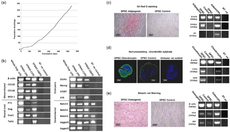
Bioactive growth factors identified within the extracellular matrix of dentine have been proposed roles in regulating the naturally inherent regenerative dentine formation seen in teeth in response to trauma and infection, which may also be harnessed for novel clinical treatments in augmenting mineralised tissue repair. This study examined the specific biological action of demineralised dentine matrix extract on a clonal population of dental pulp stem cells in stimulating the prerequisite stages of wound healing associated with mineralised tissue repair. A clonal dental pulp stem cell population with sustained proliferative capacity and multi-potentiality towards osteogenic, adipogenic and chondrogenic lineages was isolated from the pulp of human third molars. Dentine was collected from human healthy teeth, powdered and treated with ethylenediaminetetraacetic acid to obtain a solubilised DDM protein extract. The influence of DDM on the DPSC clonal population was assessed in vitro. Exposure of cells to proteolytically degraded DDM or unsupplemented media served as controls. Compared to controls, DDM stimulated cell expansion, reduced apoptotic marker caspase 3, increased cell survival marker Akt1 and enhanced mineralised matrix deposition as determined by mineral deposition and increased expression of bone-related markers, alkaline phosphatase and osteopontin. Dental pulp stem cells successfully migrated into collagen gels supplemented with demineralised dentine matrix, with cells remaining viable and expanding in numbers over a 3-day period. Collectively, the results provide evidence that soluble proteins extracted from dentine matrix are able to exert a direct biological effect on dental pulp stem cells in promoting mineralised tissue repair mechanisms.

Keywords: Dental pulp; anti-apoptotic; cell proliferation; dentine matrix; dentine repair; mesenchymal stem cells; odontogenesis; osteogenesis.


Conflict of interest statement

Declaration of conflicting interests: The authors declare that there is no conflict of interest.

Figures

Figure 1.
Figure 1.
Characterisation of the selected clonal cell population isolated from dental pulp demonstrating the isolation of an immature mesenchymal progenitor population with long proliferative lifespan and multi-potency: (a) isolated clone, designate hFNA3 achieved over 325 PDs following culture expansion; (b) RT-PCR analysis demonstrated the presence of mRNA expression for mesenchymal, neural crest, embryonic and developmental cell markers during early population doublings, which were lost at 106 PDs. Also shown is the presence of the markers in cells derived from whole dental pulp, representing a positive control; (c) demonstration for the expression of adipogenesis by Oil Red O staining and mRNA expression of early marker proliferator-activated receptor gamma (PPAR-γ) but not the later expressing marker lipoprotein lipase (LPL); (d) chondrogenesis by immunostaining for chondroitin sulphate proteoglycans and mRNA expression of type 2 collagen (Col2a) and SOX9; and (e) osteogenesis by Alizarin Red staining for mineral deposits and mRNA expression for osteocalcin (OCN) and bone sialoprotein (BSP). For all images, scale bar represents 100 µm.
Figure 2.
Figure 2.
Stimulatory effect of DDM on the expansion of hFNA3 clonal cell numbers during culture over 72 h: (a) cells were visualised by staining with crystal violet, and average cell counts and corresponding SEMs were obtained (n = 9). As a negative control, DDM was denatured to produce DDM-neg, where bioactivity was observed to be abolished, and (b) concentrations reported for DDM-neg are equivalent to the starting concentration of DDM prior to denaturation treatment. *p < 0.05, **p < 0.01 and ***p < 0.001.
Figure 3.
Figure 3.
(a) Anti-apoptotic effect of DDM on the DPSC clonal cells as demonstrated by reduced caspase 3 activity. A similar effect was not observed following supplementation with DDM-neg, and apoptosis was stimulated in the presence of the degraded products at 5 µg/mL. (b) RT-PCR for mRNA expression levels of caspase 3 and the cell survival factor Akt1 confirm the results of the caspase assay. Means and SEMs for the caspase 3 luminescence assay are calculated from n = 9. *p < 0.05, **p < 0.01 and ***p < 0.001.
Figure 4.
Figure 4.
DDM has no stimulatory or impeding influence on the migration of DPSC clonal cells into collagen gels: (a) typical confocal data derived from Z-stacks obtained from confocal microscopy images of gels indicating the ability of the cells to migrate into the collagen matrix to a depth of 50 µm; (b) collagen gels were also examined by fluorescence microscopy, with cells viewed from the top surface and cell count, using ImageJ, and are shown graphically for the respective experimental and control groups after 24 and 72 h in culture; and (c) means and SEMs are calculated from n = 15; statistical analysis indicated no significant difference between gels.
Figure 5.
Figure 5.
The ability of DDM to induce osteogenesis: (a) mRNA expression of osteogenic markers osteopontin (OPN) and alkaline phosphatase (ALP) as determined by quantitative PCR, and (b) the appearance of Alizarin Red staining mineral deposits and mineralised nodules in the presence and absence of DDM. Means and SEMs are calculated from n = 6. *p < 0.05, **p < 0.01 and ***p < 0.001.

References

    1. Smith AJ, Sloan AJ. Stem cells and the dental pulp: potential roles in dentine regeneration and repair. Oral Dis 2007; 13: 151–157. - PubMed
    1. Park ES, Cho HS, Kwon TG, et al. Proteomics analysis of human dentin reveals distinct protein expression profiles. J Proteome Res 2009; 8: 1338–1346. - PubMed
    1. Chun SY, Lee HJ, Choi YA, et al. Analysis of the soluble human tooth proteome and its ability to induce dentin/tooth regeneration. Tissue Eng Part A 2011; 17: 181–191. - PubMed
    1. Jágr M, Eckhardt A, Pataridis S, et al. Comprehensive proteomic analysis of human dentin. Eur J Oral Sci 2012; 120: 259–268. - PubMed
    1. Sloan AJ, Perry H, Matthews JB, et al. Transforming growth factor-b expression in mature human molar teeth. Histochem J 2000; 32: 247–252. - PubMed