. 2015 May 28;10(5):e0126066.
doi: 10.1371/journal.pone.0126066. eCollection 2015.

First shark from the Late Devonian (Frasnian) Gogo Formation, Western Australia sheds new light on the development of tessellated calcified cartilage

Affiliations

First shark from the Late Devonian (Frasnian) Gogo Formation, Western Australia sheds new light on the development of tessellated calcified cartilage

John A Long et al. PLoS One. .

Erratum in

Abstract

Background: Living gnathostomes (jawed vertebrates) comprise two divisions, Chondrichthyes (cartilaginous fishes, including euchondrichthyans with prismatic calcified cartilage, and extinct stem chondrichthyans) and Osteichthyes (bony fishes including tetrapods). Most of the early chondrichthyan ('shark') record is based upon isolated teeth, spines, and scales, with the oldest articulated sharks that exhibit major diagnostic characters of the group--prismatic calcified cartilage and pelvic claspers in males--being from the latest Devonian, c. 360 Mya. This paucity of information about early chondrichthyan anatomy is mainly due to their lack of endoskeletal bone and consequent low preservation potential.

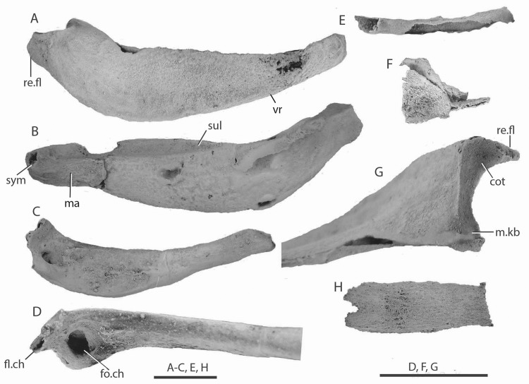
Methodology/principal findings: Here we present new data from the first well-preserved chondrichthyan fossil from the early Late Devonian (ca. 380-384 Mya) Gogo Formation Lägerstatte of Western Australia. The specimen is the first Devonian shark body fossil to be acid-prepared, revealing the endoskeletal elements as three-dimensional undistorted units: Meckel's cartilages, nasal, ceratohyal, basibranchial and possible epibranchial cartilages, plus left and right scapulocoracoids, as well as teeth and scales. This unique specimen is assigned to Gogoselachus lynnbeazleyae n. gen. n. sp.

Conclusions/significance: The Meckel's cartilages show a jaw articulation surface dominated by an expansive cotylus, and a small mandibular knob, an unusual condition for chondrichthyans. The scapulocoracoid of the new specimen shows evidence of two pectoral fin basal articulation facets, differing from the standard condition for early gnathostomes which have either one or three articulations. The tooth structure is intermediate between the 'primitive' ctenacanthiform and symmoriiform condition, and more derived forms with a euselachian-type base. Of special interest is the highly distinctive type of calcified cartilage forming the endoskeleton, comprising multiple layers of nonprismatic subpolygonal tesserae separated by a cellular matrix, interpreted as a transitional step toward the tessellated prismatic calcified cartilage that is recognized as the main diagnostic character of the chondrichthyans.


Conflict of interest statement

Competing Interests: The authors have declared that no competing interests exist.

Figures

Fig 1
Fig 1. Preparation of Gogoselachus lynbeazleyae WAM 09.6.145, Gogo Formation, Western Australia.
(A) Meckel's cartilage as exposed on collection, before acetic acid etching. (B) specimen during early acid preparation. (C) Meckel's cartilages WAM 09.6.145–001 (left), WAM 09.6.145–002 (right) after full preparation, shown articulated in dorsal view.
Fig 2
Fig 2. Head and branchial cartilages of Gogoselachus lynbeazleyae WAM 09.6.145, Gogo Formation, Western Australia.
(A–C) left Meckel’s cartilage WAM 09.6.145–001, medial, lateral and dorsal views. (D, E) ceratohyal WAM 09.6.145–005, lateral and dorsal views. (F) nasal cartilage WAM 09.6.145–006, anterior vie w?. (G)? epibranchial cartilage WAM 09.6.145–004. (H) basibranchial cartilage WAM 09.6.145–003. Abbreviations: cot, cotylus; fl.ch, flange on ceratohyal; fo.ch, fossa on ceratohyal; ma, muscle attachment area; m.kb, mandibular knob; re.fl, retroarticular flange; sul, sulcus; sym, symphyseal pit; vr, ventral ridge.
Fig 3
Fig 3. Left scapulocoracoid WAM 09.6.145–007 of Gogoselachus lynbeazleyae, Gogo Formation, Western Australia.
(A) posterior (B) medial and (C) lateral views. D, close up of posterior face showing articulation area for pectoral fin, E., interpretation of same area. F, close up of central lateral surface showing diazonal foramen (df). Abbreviations: af, articulation facets; artc, articular crest; br, break in bone; cobr, coracobrachialis muscle attachment area; cohy, coracohyoideus muscle attachment area; cp, coracoid plate; df, diazonal foramina; muscle attachment areas; flr, mediolateral pectoral retractor muscle attachment area; pdm, pectoral depressor muscle attachment area; pla, posterolateral process; pf.ar1,2, pectoral fin articulation areas 1 and 2; pla, posterolateral angle; pva, posteroventral angle; ri, ridge; sup, supinator attachment area.
Fig 4
Fig 4. Right scapulocoracoid WAM 09.6.145–007 of Gogoselachus lynbeazleyae, Gogo Formation, Western Australia.
(A) lateral (B) posterior and (C) medial views, with close up of articulatory area for pectoral fin in E, F. D, articulation area for pectoral fin articulation on left scapulocoracoid for comparison. Abbreviations as for Fig 3.
Fig 5
Fig 5. Teeth of Gogoselachus lynbeazleyae WAM 09.6.145, Gogo Formation, Western Australia.
Large tooth WAM 09.6.145–009 in (A) lingual, (B) labial, (C) oral (D) aboral views, in natural light. (E) SEM of tooth WAM 09.6.145–010, naturally broken near the median surface. (F, G) CT-scans of tooth WAM 09.6.145–011, showing layout of vascular canals, in basal and lingual-lateral views. Abbreviations: l-lcl, labio-lingual canals; ctcl, central canal; blcl,basolabial canals; ccl, coronal canals.
Fig 6
Fig 6. SEM images of teeth of Gogoselachus lynbeazleyae WAM 09.6.145, Gogo Formation, Western Australia.
(A–C) WAM 09.6.145–012 in oral, labial, and lingual views. (D–F) WAM 09.6.145–013 in labial, basal, and oblique lateral views. (G–H) Small tooth, WAM 09.6.145–014, in lingual, and oral views. (I) Tooth of intermediate size, WAM 09.6.145–015, in labial view. (J–M) WAM 09.6.145–016 in lingual, labial, oral and distal views. (N–P) WAM 09.6.145–017 in oral, labial, and lingual views. (Q–T) WAM 09.6.145–018 in labial, lingual, oral, and lateral views.
Fig 7
Fig 7. SEM images of scales of Gogoselachus lynbeazleyae WAM 09.6.145, Gogo Formation, Western Australia.
(A) presumed flank scale WAM 09.6.145–019, anterocrown view. (B, C) presumed flank scale WAM 09.6.145–020, anterocrown and posterior views. (D) presumed flank scale WAM 09.6.145–021, posterobasal view. (E, F) presumed flank scale WAM 09.6.145–022, posterobasal view and magnification of undersurface of crown denticulation. (G, H) very small scale WAM 09.6.145–023, broken, showing crown view of one half and basal view of the other half. (I) abraded umbellate scale WAM 09.6.145–024, crown view. (J, K) Pultschuppe or squamae proniae scale WAM 09.6.145–025, crown and posterior views. (L) stellate tessera WAM 09.6.145–026, crown view. (M) probable branchial denticle plate WAM 09.6.145–027, lateral view. (N) vertical transverse section through presumed flank scale WAM 09.6.145–028. (O) oblique vertical section through umbellate scale WAM 09.6.145–029. (P, Q) horizontal section through the crown of? head tessera WAM 09.6.145–030, whole section and magnification to show Spiralfasern structure. Scale bars = 0.1 mm in all figures except (P), where scale bar = 1 mm.
Fig 8
Fig 8. Structure of Gogoselachus lynbeazleyae endoskeleton.
(A–C) Gogoselachus calcified cartilage (A) SEM WAM 09.6.145–031 showing tessellate layout (B, C) horizontal section through tissue WAM 09.6.145–032 (D) transverse CT scan of right Meckel's cartilage. Scale bar = 0.1 mm in (C). Abbreviations: cp, cell processes; l, lacunae.
Fig 9
Fig 9. Comparative endoskeletal tissues in other Devonian gnathostomes.
(A) globular (or granular) calcified cartilage preserved in the head region on stem chondrichthyan Doliodus problematicus NBMG 10127. (B) vertical thin section of large calcified cartilage globules in Euthacanthus macnicoli NMS G.2010.7.40. (C–E) placoderm endoskeletal tissues (C) SEM of QMF53545, perichondral bone sheet with honeycomb ‘cells’ in an antiarch from the upper Lower Devonian Cravens Peak Beds, western Queensland. (D) scans of perichondral bone from the Early Devonian Parabuchanosteus murrumbidgeensis from Taemas, ANU V24x. (E) section of sheet of crystalline calcite associated with a specimen of antiarch placoderm Bothriolepis canadensis; arrows show separation between individual crystals. (F–H) prismatic calcified cartilage of an undetermined shark from the Early Carboniferous Laurel Formation, northwestern Australia, QMF57586, QMF57587. (F) transmitted light image of QMF57588 showing variation in tesserae size (G, H) vertical section showing layout of tesserae, with prismatic structure visible under cross-nicols in (H). Scale bars = 1 mm in (A), (F), 0.1 mm in (B), (E), (G).
Fig 10
Fig 10. Teeth of sharks from the Upper Devonian of central Iran.
(A–C) Arduodens flammeus Hairapetian & Ginter, 2009, from Chahriseh, AEU 610, in lingual, aboral, and lateral views. (D–G) Deihim mansureae Ginter, Hairapetian and Klug, 2002. (D, E) Holotype, IGPUW/Ps/5/1, from Hutk, in oral and lateral views. (F, G) Putative anterior tooth, AEU 239, from Hodjedk, in labial and oral views. Scale bar = 0.5 mm.

References

    1. Schaeffer B (1975) Comments on the origin and basic radiation of the gnathostome fishes with particular reference to the feeding mechanism In: Lehman J- P, editor. Problèmes actuels de paléontologie: evolution des Vertébrés. Paris: Colloques Internationaux du Centre National de la Recherche Scientifique; pp. 101–109.
    1. Janvier P (1996) Early Vertebrates. Oxford: Oxford University Press. 393 p.
    1. Brazeau M (2009) The braincase and jaws of a Devonian ‘acanthodian’ and modern gnathostome origins. Nature 457: 305–308. 10.1038/nature07436 - DOI - PubMed
    1. Davis SP, Finarelli JA, Coates MI (2012) Acanthodes and shark-like conditions in the last common ancestor of modern gnathostomes. Nature 486: 247–250. 10.1038/nature11080 - DOI - PubMed
    1. Zhu M, Yu XB, Ahlberg PE, Choo B, Lu J, et al. (2013) A Silurian placoderm with osteichthyan-like marginal jaw bones. Nature 502: 188–193. 10.1038/nature12617 - DOI - PubMed

Publication types

LinkOut - more resources