Review
. 2015 Sep;23(9):558-66.
doi: 10.1016/j.tim.2015.04.010. Epub 2015 May 25.

Bacterial spread from cell to cell: beyond actin-based motility

Affiliations
Review

Bacterial spread from cell to cell: beyond actin-based motility

Carole J Kuehl et al. Trends Microbiol. 2015 Sep.

Abstract

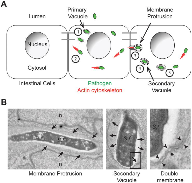
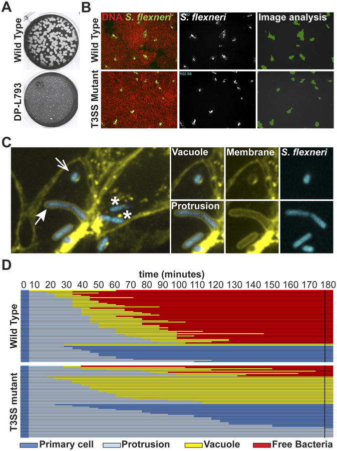
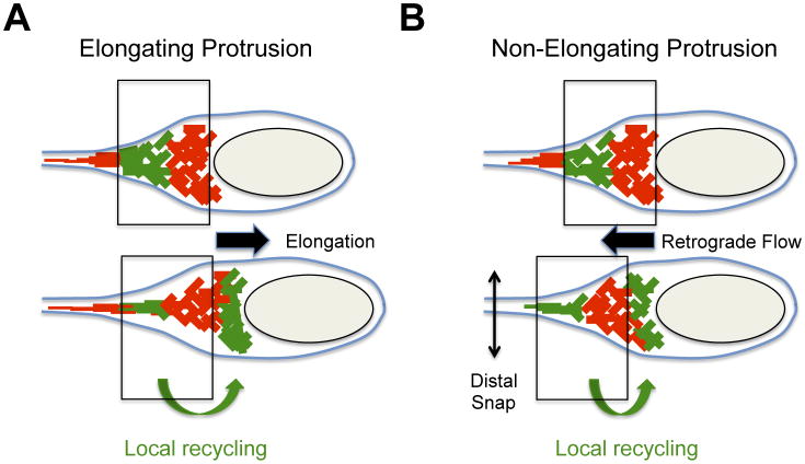
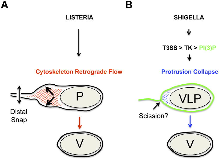
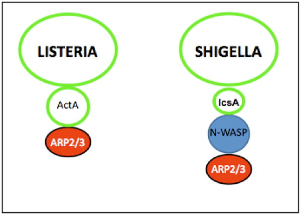
Several intracellular pathogens display the ability to propagate within host tissues by displaying actin-based motility in the cytosol of infected cells. As motile bacteria reach cell-cell contacts they form plasma membrane protrusions that project into adjacent cells and resolve into vacuoles from which the pathogen escapes, thereby achieving spread from cell to cell. Seminal studies have defined the bacterial and cellular factors that support actin-based motility. By contrast, the mechanisms supporting the formation of protrusions and their resolution into vacuoles have remained elusive. Here, we review recent advances in the field showing that Listeria monocytogenes and Shigella flexneri have evolved pathogen-specific mechanisms of bacterial spread from cell to cell.

Keywords: Listeria monocytogenes; Shigella flexneri; double-membrane vacuole; membrane protrusion; spread from cell to cell.


Figures

Figure 1
Figure 1. Sequence of events in bacterial spread from cell to cell
(A) Cytosolic bacteria (green) spread from cell to cell within a monolayer of intestinal cells through the following sequence of events: (1) Escape from the primary vacuole, (2) Actin (red)-based motility, (3) Membrane protrusion formation into adjacent cells, (4) Resolution of membrane protrusions into (double-membrane) secondary vacuoles and (5) Escape from secondary vacuoles into the cytosol of the adjacent cell. Adapted from reference [1]. (B) Electron micrographs of the two main features of bacterial cell-to-cell spread, membrane protrusions and double membrane vacuoles. Left panel: S. flexneri (S.f) within a membrane protrusion in between two lobes of the adjacent cell nucleus (n). Membranes surrounding the protrusion are marked with arrows. Middle panel: S. flexneri within a secondary vacuole. Membranes surrounding the secondary vacuoles are marked with arrows. Right panel: high magnification showing the double membranes of a secondary vacuole corresponding to the boxed area in the middle panel. Double membranes are marked with opposing arrowheads.
Figure 2
Figure 2. Readouts of bacterial spread from cell to cell
(A) L. monocytogenes plaque assay depicting large (wild type) plaques, and a small plaque phenotype (DP-L793). Adapted from reference [7]. (B) Automated microscopy of S. flexneri infection in HT-29 cell monolayer. Left panels, overlay of DNA staining (red) and GFP (green)-expressing wild type and type III secretion system (T3SS) mutant strains. Middle panels: bacterial infection foci. Right panels: computer-assisted image analysis of infection foci size (green). (C) High-magnification confocal microscopy showing features of bacterial spread form cell to cell in HT-29 cells expressing a fluorescent membrane marker (yellow) infected with S. flexneri (blue). Cytosolic bacteria in a primary infected cell (*) form membrane protrusions (closed arrow) that project into adjacent cells and resolve into secondary vacuoles (open arrow). (D) Tracking analysis of 60 wild type (top) and T3SS mutant (bottom) strains. Each line represents one bacterium that was tracked for 180 minutes and the progression of the dissemination process is depicted using the color key shown at the bottom. Primary cell, dark blue; Protrusion, light blue; Vacuole, yellow; Free bacteria in adjacent cell, red. The data shows that 75% of the wild type bacteria succeed in forming protrusions that resolve into vacuoles from which the bacteria escape (free). By contrast, the majority of the T3SS mutant bacteria either form protrusions that fail to resolve into vacuoles and retract to the primary infected cell, or fails to escape the formed vacuole. Adapted from reference [18].
Figure 3
Figure 3. Dynamics of the actin cytoskeleton in protrusions
Dynamics of the actin cytoskeleton were determined by photo-activation experiments with photo-activatable GFP-actin fusion protein. Photo-activated GFP-actin (green network) demonstrating local recycling (green arrow at the bottom) from the distal network (top panel) to the bacterial pole (bottom panel). The vertical boxes indicate the position of the actin network at the bacterial pole at the instant of photo-activation (top panel) and shortly after photo-activation (bottom panel). (A) Cytoskeleton dynamics in elongating protrusions. The disassembly of the distal network (top panel, photo-activated actin-GFP, green) fuels the assembly of the proximal network at the bacterial pole (bottom panel, photo-activated actin-GFP, green). Note that the network at the bacterial pole did not move with reference to the plasma membrane (vertical boxes). The assembly of the network at the bacterial pole provides the forces leading to protrusion elongation (black arrow). (B) Cytoskeleton dynamics in non-elongating protrusions. The disassembly of the distal network (top panel, photo-activated actin-GFP, green) fuels the assembly of the proximal network at the bacterial pole (bottom panel, photo-activated actin-GFP, green). Note that the network at the bacterial pole moves backwards (retrograde flow) with reference to the plasma membrane (vertical boxes). The assembly of the network at the bacterial pole provides the forces leading to retrograde flow (black arrow) and membrane scission (distal snap).
Figure 4
Figure 4. Mechanisms of L. monocytogenes and S. flexneri protrusion resolution
(A) Mechanism of L. monocytogenes protrusion resolution. After elongation, the actin network (red lines) in protrusions (P) undergoes retrograde flow (black arrow), which generates the forces leading to membrane scission (vertical arrow, distal snap) and vacuole formation (V). (B) Mechanism of S. flexneri protrusion resolution. After elongation, the actin network (blue lines) collapses in response to PI(3)P production in the protrusion membrane (green line). PI(3)P production is mediated by a signaling cascade involving the type 3 secretion system (T3SS), host cell tyrosine kinase (TK) and PI3KC2A-dependent production of PI(3)P. Cytoskeleton collapse leads to the formation of vacuole-like protrusions (VLP) and the resolution of the membrane tether (scission?) connecting VLP to the primary infected cells leads to the formation of a genuine vacuole (V).
Figure I
Figure I
Bacterial and cellular factors supporting Listeria monocytogenes and Shigella flexneri actin-based motility.

References

    1. Tilney LG, Portnoy DA. Actin filaments and the growth, movement, and spread of the intracellular bacterial parasite, Listeria monocytogenes. J Cell Biol. 1989;109:1597–1608. - PMC - PubMed
    1. Gouin E, Gantelet H, Egile C, Lasa I, Ohayon H, et al. A comparative study of the actin-based motilities of the pathogenic bacteria Listeria monocytogenes, Shigella flexneri and Rickettsia conorii. J Cell Sci. 1999;112(Pt 11):1697–1708. - PubMed
    1. Kadurugamuwa JL, Rohde M, Wehland J, Timmis KN. Intercellular spread of Shigella flexneri through a monolayer mediated by membranous protrusions and associated with reorganization of the cytoskeletal protein vinculin. Infect Immun. 1991;59:3463–3471. - PMC - PubMed
    1. Monack DM, Theriot JA. Actin-based motility is sufficient for bacterial membrane protrusion formation and host cell uptake. Cell Microbiol. 2001;3:633–647. - PubMed
    1. Takeuchi A, Formal SB, Sprinz H. Exerimental acute colitis in the rhesus monkey following peroral infection with Shigella flexneri. An electron microscope study. Am J Pathol. 1968;52:503–529. - PMC - PubMed

Publication types