. 2015 Nov;25(11):4572-83.
doi: 10.1093/cercor/bhv098. Epub 2015 May 28.

Serotonin Attenuates Feedback Excitation onto O-LM Interneurons

Affiliations

Serotonin Attenuates Feedback Excitation onto O-LM Interneurons

Claudia Böhm et al. Cereb Cortex. 2015 Nov.

Abstract

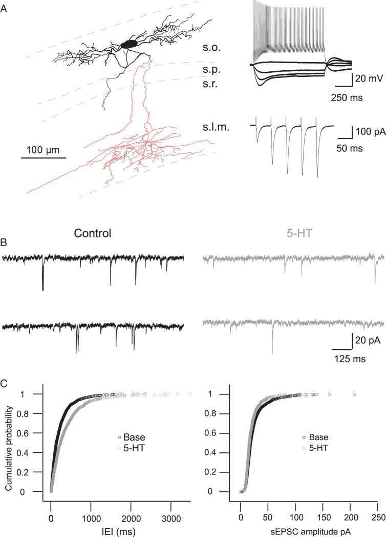
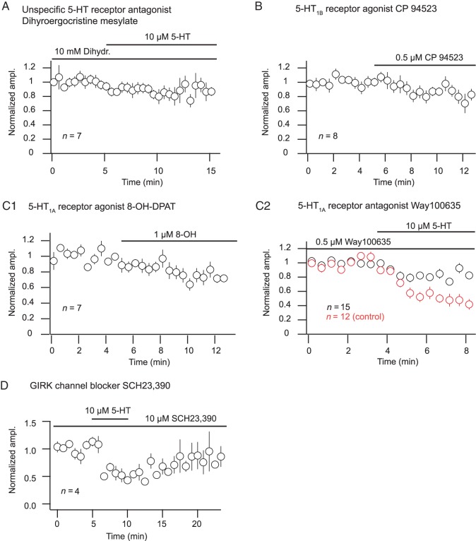
The serotonergic system is a subcortical neuromodulatory center that controls cortical information processing in a state-dependent manner. In the hippocampus, serotonin (5-HT) is released by ascending serotonergic fibers from the midbrain raphe nuclei, thereby mediating numerous modulatory functions on various neuronal subtypes. Here, we focus on the neuromodulatory effects of 5-HT on GABAergic inhibitory oriens lacunosum-moleculare (O-LM) cells in the hippocampal area CA1 of the rat. These interneurons are thought to receive primarily local excitatory input and are, via their axonal projections to stratum lacunosum-moleculare, ideally suited to control entorhinal cortex input. We show that 5-HT reduces excitatory glutamatergic transmission onto O-LM interneurons. By means of paired recordings from synaptically connected CA1 pyramidal cells and O-LM interneurons we reveal that this synapse is modulated by 5-HT. Furthermore, we demonstrate that the reduction of glutamatergic transmission by serotonin is likely to be mediated via a decrease of calcium influx into presynaptic terminals of CA1 pyramidal cells. This modulation of excitatory synaptic transmission onto O-LM interneurons by 5-HT might be a mechanism to vary the activation of O-LM interneurons during ongoing network activity and serve as a brain state-dependent switch gating the efficiency of entorhinal cortex input to CA1 pyramidal neurons.

Keywords: 5-HT; CA1; paired recordings; presynaptic neuromodulation.


Figures

Figure 1.
Figure 1.
5-HT inhibits spontaneous EPSCs in O-LM interneurons. (A) Left, reconstruction of an O-LM interneuron (dashed: outline of cell layers, black: cell body and dendrites, red: axon). Right top, typical voltage responses of an O-LM cell to de- and hyperpolarizing current pulses. Right bottom, strongly facilitating excitatory postsynaptic current amplitudes to consecutive extrasynaptic stimuli. (B) Example traces of spontaneous EPSCs under control conditions and in 10 µM 5-HT. (C) Cumulative probability of interevent intervals (IEI, left) and amplitude of spontaneous EPSCs (right) under control conditions, in 10 µM 5-HT and after wash (n = 8). s.l.m., stratum lacunosom-moleculare; s.o., stratum oriens, s.p., stratum pyramidale; s.r., stratum radiatum.
Figure 2.
Figure 2.
Bath-applied as well as endogenously released 5-HT decreases stimulus-evoked EPSCs. (A) Recording configuration with an extracellular stimulating electrode positioned at the border of the alveus and stratum oriens. (B1) Top, example traces of stimulus-evoked EPSCs. Bottom, time course of the EPSC amplitude before, during and after washout of 10 µM 5-HT. (B2) Summary of the time course of the normalized and binned EPSC amplitudes (n = 12). (C1) Top, example traces of stimulus-evoked EPSCs. Bottom, time course of the EPSC amplitude before, during and after washout of 200 µM fenfluramine. (C2) Summary of time course of the normalized and binned EPSC amplitudes (n = 7). (D) Endogenously released 5-HT reduces spiking probability. (D1) Example experiment where spikes were evoked in an O-LM interneuron by extracellular stimulation. Five pulses at theta frequency (interstimulus interval: 125 ms) were delivered. * Indicate stimulus time points. Top, example traces in control conditions (black) and in fenfluramine (200 µM, gray). (Inset) EPSP evoked by the first stimulus in control condition (black) and in fenfluramine (gray), average of 4 traces each, scale bar, x: 20 ms, y: 5 mV. Bottom, time course of experiment displayed as raster plot of spikes evoked in response to stimulation. Spikes were abolished shortly after washing in fenfluramine (gray area). (D2) Summary, spike count normalized to the summed and averaged spike counts of trials under control conditions. Inset, averaged spike counts under control conditions and in fenfluramine (n = 5).
Figure 3.
Figure 3.
Serotonin reduces glutamatergic excitatory synaptic transmission at the CA1 pyramidal cell–O-LM interneuron synapse. (A) Reconstruction of a synaptically connected pyramidal cell–O-LM interneuron pair (dashed: outline of cell layers, black: cell body and dendrites of O-LM interneuron, red: axon of O-LM interneuron; gray: cell body and dendrites of pyramidal cell, blue: axon of pyramidal cell). Middle, left, voltage responses of the shown O-LM cell to de- and hyperpolarizing current pulses. (B1) Strongly facilitating unitary excitatory postsynaptic currents to consecutive APs, elicited in the presynaptic pyramidal cell. Below the postsynaptic response to consecutive APs after 5-HT application and after washout of 5-HT are shown. (B2) Grand average of the postsynaptic responses of 4 connected pairs in the indicated conditions. (C) Traces of uEPSCs evoked by the first presynaptic AP (time point indicated by arrow) in 4 cells under control conditions, after application of 10 µM 5-HT and after washout of 5-HT. (D) Summary of the reduction of the normalized first uEPSC amplitude by application of 5-HT and after washout of 5-HT (n = 4). s.l.m., stratum lacunosom-moleculare; s.o., stratum oriens, s.p., stratum pyramidale; s.r., stratum radiatum.
Figure 4.
Figure 4.
5-HT acts most likely presynaptically. (A) Left, example traces of miniature EPSCs in control conditions (black) and in 10 µM 5-HT (gray). Right, cumulative probability of interevent intervals and amplitude of miniature EPSCs in control conditions and in 10 µM 5-HT (n = 10). (B) Top, recording configuration for glutamate uncaging. With a laser flash glutamate is uncaged in the immediate vicinity of the soma of an O-LM interneuron. Middle, example traces of the glutamate evoked current before (black) and after the application of 10 µM 5-HT (gray). Bottom, summary of the time course of the glutamate evoked current. Depicted is the normalized and binned peak amplitude. Note that the amplitude is not decreased after application of 5-HT (n = 4). (C) Left, example of connected pyramidal–O-LM pair, top, overlay of uEPSCs in control conditions and in 5-HT, middle, time course of successful synaptic transmission and failures. Bottom, time course of the uEPSC amplitude (as shown in top row) before, during and after washout of 10 µM 5-HT. Note that the failure rate increases during application of 5-HT. Right, summary of failure rate in synaptically connected paired recordings under control conditions and in 10 µM 5-HT (n = 4).
Figure 5.
Figure 5.
Effect of 5-HT receptor subtype (un)specific compounds on eEPSC amplitude. (AD) Time course of averaged, normalized, and binned amplitude. The paired Wilcoxon sign rank test was used to compare amplitudes in the indicated conditions (tested on raw, not normalized amplitudes). (A) In the presence of the unspecific 5-HT receptor antagonist dihydroergocristine mesylate the effect of 5-HT on the EPSC amplitude is abolished. (n = 7, P = 0.30). (B) The 5-HT1B receptor agonist CP 94523 does not mimic the 5-HT effect. (n = 8, P = 0.15). (C1) The 5-HT1A receptor agonist 8-OH-DPAT reduces eEPSC amplitudes (n = 7, P = 0.02). (C2) The 5-HT1A receptor antagonist WAY100635 partially blocks the 5-HT effect on eEPSC amplitude (n = 15, P = 0.01). For comparison the eEPSC amplitude in control conditions, that is, in the absence of the antagonist is shown (n = 12) (red, compare Fig. 2B, unpaired Wilcoxon test, P = 0.0002). (D) In the presence of the GIRK-channel blocker SCH23390 5-HT still reduces the amplitude of eEPSCs (n = 4). Also compare Figure 2B2.
Figure 6.
Figure 6.
Decreased presynaptic calcium influx can account for the reduction of glutamatergic transmission by 5-HT. (A) Ca2+-imaging recording configuration. Axonal fibers were filled with the Ca2+-sensitive dye magnesium green AM (green) by bulk loading and stimulated with an extracellular electrode positioned at the border of the alveus and stratum oriens. The resulting Ca2+ transients were measured with a photodiode. (B) Top, example traces of the Ca2+ transients in control and in 20 µM CdCl2. Bottom, time course of ΔF/F amplitude of the Ca2+ transients shown above. Note that the transient is abolished in the presence of the calcium channel blocker CdCl2. (C) Top, example traces of the calcium transient in control, in 10 µM 5-HT and after wash. Bottom, time course of ΔF/F amplitude of the above transients. (D) Summary of time course of ΔF/F amplitude (n = 11).
Figure 7.
Figure 7.
The 5-HT induced reduction of the presynaptic Ca2+ transient as well as the reduction of the glutamatergic synaptic transmission can be mimicked by a reduction of the extracellular Ca2+ concentration. (A) Top, example traces of Ca2+ signals in control conditions, after application of 10 µM 5-HT, after washout of 5-HT and after reduction of the extracellular Ca2+ concentration from 2.5 to 2 mM. Bottom, time course of the binned amplitude of the Ca2+ transient in control conditions, after application of 10 µM 5-HT, after washout and in 2 mM Ca2+. (B) Example traces of stimulus-evoked EPSCs in control conditions and after reduction of the extracellular Ca2+ concentration from 2.5 to 2 mM. (C) Summaries of the effect of 5-HT and the reduced extracellular Ca2+ concentration on the amplitude of the Ca2+ transients and on the amplitude of stimulus-evoked EPSCs.

References

    1. Ali AB, Thomson AM. 1998. Facilitating pyramid to horizontal oriens-alveus interneurone inputs: dual intracellular recordings in slices of rat hippocampus. J Physiol (Lond). 507(Pt 1):185–199. - PMC - PubMed
    1. Andrade R, Nicoll RA. 1987. Pharmacologically distinct actions of serotonin on single pyramidal neurones of the rat hippocampus recorded in vitro. J Physiol (Lond). 394:99–124. - PMC - PubMed
    1. Araque A, Parpura V, Sanzgiri RP, Haydon PG. 1999. Tripartite synapses: glia, the unacknowledged partner. Trends Neurosci. 22:208–215. - PubMed
    1. Barnes NM, Sharp T. 1999. A review of central 5-HT receptors and their function. Neuropharmacology. 38:1083–1152. - PubMed
    1. Biró AA, Holderith NB, Nusser Z. 2005. Quantal size is independent of the release probability at hippocampal excitatory synapses. J Neurosci. 25:223–232. - PMC - PubMed

Publication types

MeSH terms