Clinical Trial
. 2015 May 29;6(1):110.
doi: 10.1186/s13287-015-0105-2.

Efficacy of autologous stem cell-based therapy for osteonecrosis of the femoral head in sickle cell disease: a five-year follow-up study

Affiliations
Clinical Trial

Efficacy of autologous stem cell-based therapy for osteonecrosis of the femoral head in sickle cell disease: a five-year follow-up study

Gildasio Cerqueira Daltro et al. Stem Cell Res Ther. .

Abstract

Introduction: Stem cell therapy with bone marrow-derived mononuclear cells (BMMCs) is an option for improving joint function in osteonecrosis of the femoral head (ONFH). Bone marrow-derived mesenchymal stromal cell (MSC) numbers and their osteogenic differentiation are decreased in patients with ONFH. However, whether this decrease also extends to the early stages of ONFH in sickle cell disease (SCD) is still unclear.

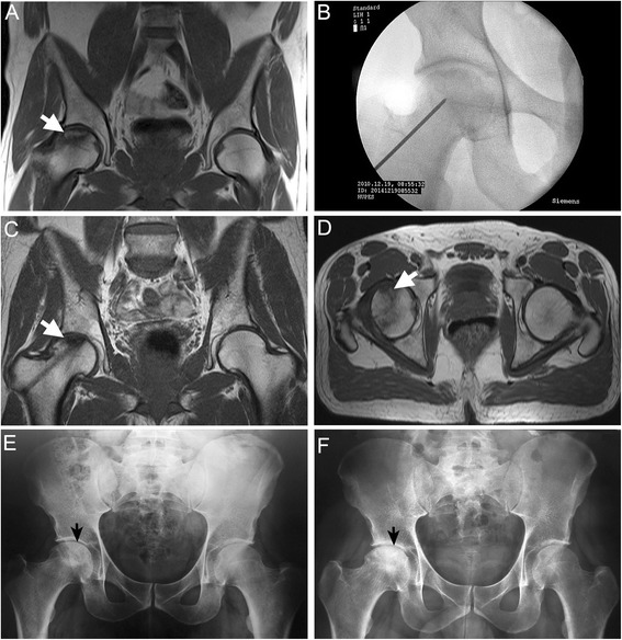
Methods: We conducted a phase I/II, non-controlled study to determine efficacy and safety of BMMC implantation using a minimally invasive technique in SCD patients with ONFH. Eighty-nine patients were recruited and followed up for 60 months after surgery. Clinical and radiographic findings were assessed, and data were completed by in vitro analysis.

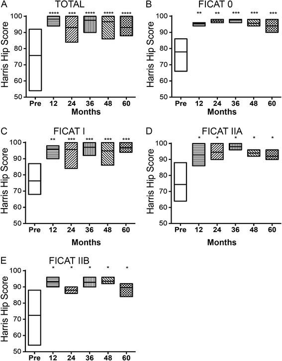
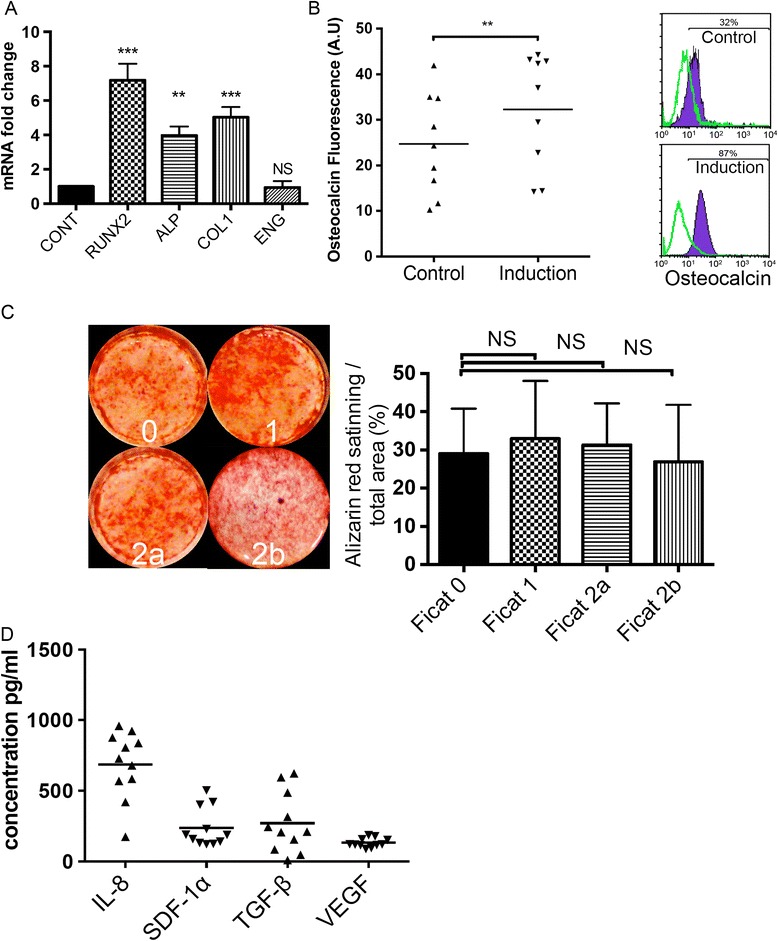
Results: At the final follow-up (60 months) there was a significant improvement in clinical joint symptoms and pain relief as measured by the Harris Hip Score (P = 0.0005). In addition, after the BMMC implantation procedure, radiographic assessment showed disease stabilization and only 3.7 % of the treated patients did not achieve a satisfactory clinical result. The amount of fibroblast colony-forming units was 28.2 ± 13.9 per 1 million BMMCs after concentration. Flow cytometry analysis showed a significantly higher number of hematopoietic stem/endothelial progenitor cell markers in concentrated BMMCs when compared with bone marrow aspirate, indicating an enrichment of these cell types. Isolated MSCs from SCD patients with pre-collapse ONFH maintained the replicative capacity without significant loss of their specific biomolecular characteristics, multi-differentiation potential, and osteogenic differentiation activities. Cytokines and growth factors (interleukin-8, transforming growth factor-beta, stromal cell-derived factor-1alpha and vascular endothelial growth factor) that mediate endogenous bone regeneration were also produced by expanded MSCs from SCD patients.

Conclusion: The autologous BMMC implantation with a minimally invasive technique resulted in significant pain relief and halted the progression of early stages of ONFH in SCD patients. MSCs from SCD patients display biological properties that may add to the efficiency of surgical treatment in ONFH. In summary, our results indicate that infusion of BMMCs enriched with stem/progenitor cells is a safe and effective treatment for the early stages of ONFH in SCD patients.

Trial registration: ClinicalTrials.gov NCT02448121; registered 15 May 2015.


Figures

Fig. 1
Fig. 1
BMMC treatment significantly improved the outcome of femoral heads at early-stage ONFH in SCD. ae Changes in HHS for femoral heads grouped according to Ficat stage over the entire postoperative period. Floating bars represent maximum and minimum range of variation, with lines at mean HHS. *P < 0.05, **P < 0.01, ***P < 0.001, ****P < 0.0001, versus preoperative HHS values
Fig. 2
Fig. 2
Radiographic sequence from a representative case of a symptomatic 44-year-old man with ONFH successfully treated with BMMC implantation. a Coronal T1-weighted MRI scans of the right hip before surgery show osteonecrotic lesion in Ficat IIB stage. The osteonecrotic lesion is surrounded by sclerotic low-signal margin (arrow) with subjacent marrow edema. b Anteroposterior X-ray radiograph showing minimally invasive decompression. The trephine was introduced under fluoroscopy through the greater trochanter into the femoral head. The BMMCs were injected through the trephine into the necrotic zone. c, d Twelve-month postoperative coronal (c) and axial (d) MRI (T1-weighted) scans after autologous BMMC implantation showing the channel, decreased bone edema and changes of signal in the superior part of the femur. Axial MR image (d) shows well-delimited marrow region of increased signal intensity (arrow) within the necrotic area and hypointense line around them due to reparative new bone. e, f Preoperative (e) and 60-month postoperative anteroposterior (f) radiographs of the same hip showing the femoral head has maintained the sphericity (arrow)
Fig. 3
Fig. 3
Increased number of conjunctive and osteogenic progenitor cells in concentrated BMMC. a Scatter plots showing the distribution of total mononuclear cell number in 1 × 10⁶ cells/mL after bone marrow aspiration (BMA) and after concentration of bone marrow-derived mononuclear cells (BMMC) on Ficoll density gradient centrifugation. b, c Flask and morphologic characteristics of colony-forming unit-fibroblasts (CFU-F) from BMMC. d, e Flask with mineralized matrix stained with red Alizarin-S and Crystal Violet of colony-forming unit-osteoblasts (CFU-O) from BMMC. f Frequency of CFU-F/106 cells and CFU-O/106 cells in concentrated BMMC. g Frequency of CFU-F/106 cells isolated in BMMC from individuals with osteonecrosis at a distant site was independent of the disease stage. The high standard deviation indicates the high variability of CFU-F and CFU-O within the SCD bone marrow. Data are represented as individuals values. Horizontal bars indicate mean value. Scale bar: 50 μm. ***P < 0.001. NS, not significant
Fig. 4
Fig. 4
Concentrated number of endothelial and hematopoietic progenitor cell phenotypes in BMMC. a–d The frequency of cell subpopulations was obtained after labeling of total bone marrow-derived mononuclear cells (BMMC) (1 × 10⁶ cells) separately with antibodies (set 1 CD133/CD34/KDR or set 2 CD34/KDR/CD45) and analysis by flow cytometry. Phenotypes CD34+/KDR+ (a), CD34+/KDR+/CD133+ (b), CD34/CD45dim (c) and CD34+/KDR+/CD45dim (d) were concentrated in the implanted BMMC of SCD patients. e Concentration factor calculated by the quotient of the specified phenotype number in BMMC/BMA. The high standard deviation indicates the high variability of cell phenotypes within the SCD bone marrow. Data are represented as individuals values. Horizontal bars indicate mean value. *P < 0.05, **P < 0.01, ***P < 0.005, ****P < 0.001. BMA bone marrow aspirate
Fig. 5
Fig. 5
Expansion, characterization and pluripotency properties of human MSCs from SCD patients. a Ex vivo expanded mesenchymal stromal cells (MSC) from SCD patients with pre-collapse ONFH at a distant site in control culture conditions. Total viable cell counts of MSCs from donors with progressive disease stage are plotted for MSCs expanded in fetal bovine serum. b Morphologic characteristics of MSCs at passage 3. ce Differentiation of MSCs. Results of staining assays on MSCs induced to osteogenic differentiation (c, mineralized bone nodules stained with Alizarin red), adipogenic differentiation (d, intracellular lipid droplets stained with Oil red O), and chondrogenic differentiation (e, static micromass cell culture stained with Alcian Blue). f Immunophenotyping profile of MSCs isolated from SCD patients. Negative peaks (green line) correspond to cell autofluorescence. MSCs were positively labeled to CD29, CD90, CD105, CD146 and negatively labeled to CD31, CD34, CD45 and CD133. Data are mean ± SD number of cells (n = 4). Data are representative of three independent experiments. Magnification: 400× in b–d. Scale bar: 50 μm in e
Fig. 6
Fig. 6
Osteogenic abilities of bone marrow-derived MSCs are not defective in patients with osteonecrosis. a Quantitative RT-PCR of osteoblast-related genes from control and osteogenic-induced MSCs after 10 days were normalized and compared according to housekeeping genes as control. b Scatter plots (left) showing the distribution of total fluorescence intensity from FACS analysis (right) of cellular osteocalcin expression in MSCs. The relative fluorescence intensity shows significant differences between control and osteogenic differentiation conditions. c Alizarin Red staining (left) indicating mineralized nodule formation of cultured MSCs after treatment with osteogenic media for 21 days. Quantitative analysis of the amount of Alizarin staining in each group is also indicated (right). No significant difference (NS) was found between MSCs from SCD patients with early-stage osteonecrosis and patients with further stages of the disease at a distant site. Alizarin Red staining was independent of the disease stage. d Enzyme-linked immunosorbent assay for interleukin-8 (IL-8), transforming growth factor-beta (TGF-β), stromal cell-derived factor-1alpha (SDF-1α) and vascular endothelial growth factor (VEGF). Concentration (pg/mL) of cytokines and growth factors in supernatants of MSCs of SCD patients after 2 days of culture. MSCs from SCD patients produce distinct levels of IL-8. Horizontal bars represent the mean and SD of data. The triangles represent individual values. *P < 0.05, **P < 0.01, ***P < 0.001

References

    1. Adorno EV, Couto FD, Moura Neto JP, Menezes JF, Rego M, Reis MG, et al. Hemoglobinopathies in newborns from Salvador, Bahia, Northeast Brazil. Cad Saude Publica. 2005;21:292–8. doi: 10.1590/S0102-311X2005000100032. - DOI - PubMed
    1. Adorno EV, Zanette A, Lyra I, Seixas MO, Reis MG, Gonçalves MS. Clinical and molecular characteristics of sickle cell anemia in the northeast of Brazil. Genet Mol Biol. 2008;31:621–5. doi: 10.1590/S1415-47572008000400003. - DOI
    1. Gonçalves MS, Queiroz IL, Cardoso SA, Zanetti A, Strapazoni AC, Adorno E, et al. Interleukin 8 as a vaso-occlusive marker in Brazilian patients with sickle cell disease. Braz J Med Biol Res. 2001;34:1309–13. doi: 10.1590/S0100-879X2001001000011. - DOI - PubMed
    1. Acurio MT, Friedman RJ. Hip arthroplasty in patients with sickle-cell haemoglobinopathy. J Bone Joint Surg (Br) 1992;74:367–71. - PubMed
    1. Kato GJ, Gladwin MT, Steinberg MH. Deconstructing sickle cell disease: reappraisal of the role of hemolysis in the development of clinical subphenotypes. Blood Rev. 2007;21:37–47. doi: 10.1016/j.blre.2006.07.001. - DOI - PMC - PubMed

Publication types

MeSH terms

Associated data