. 2015 May 29:5:10779.
doi: 10.1038/srep10779.

Oocyte quality is decreased in women with minimal or mild endometriosis

Affiliations

Oocyte quality is decreased in women with minimal or mild endometriosis

Bo Xu et al. Sci Rep. .

Abstract

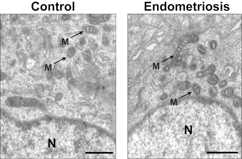
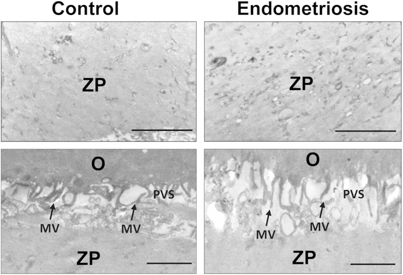
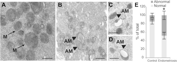
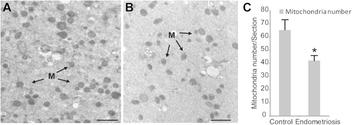
Endometriosis, a pathological condition in which the endometrium grows outside the uterus, is one of the most common causes of female infertility; it is diagnosed in 25-40% of infertile women. The mechanism by which endometriosis affects the fertility of females remains largely unknown. We examined the ultrastructure of oocytes from patients with minimal or mild endometriosis and control females undergoing in vitro fertilization (IVF) treatment by transmission electron microscopy (TEM) to investigate the physiological significance of oocyte quality for patients with minimal or mild endometriosis. The TEM results revealed that the oocytes from women with minimal or mild endometriosis exhibited abnormal mitochondrial structure and decreased mitochondria mass. Quantitative real time PCR analysis revealed that the mitochondrial DNA copy number was significantly reduced in the oocytes from women with minimal or mild endometriosis compared with those of the control subjects. Our results suggest that decreased oocyte quality because of impaired mitochondrial structure and functions probably an important factor affecting the fertility of endometriosis patients.


Figures

Figure 1
Figure 1. Cumulus cell of the oocytes in the endometriosis and control groups.
The cumulus cells from the control group show the same ultrastructural cytoplasmic characteristics of the cumulus cells surrounding the oocytesas in the endometriosis group (A,B). The tubular cristae of the mitochondria (arrows) are well developed and evenly distributed. N = nuclei; M = mitochondria; Scale bar (A,B) = 500 nm.
Figure 2
Figure 2. The electron density of the zona pellucida (A,B) and perivitelline space (C,D) in the endometriosis and control groups, respectively.
No difference was observed between the endometriosis and control groups in the dense appearance of the inner aspect of the ZP, and some fibres are visible in the zona texture (A,B). The microvilli (arrows) are numerous and long on the oolemma of both groups (C,D). MV = microvilli; PVS = perivitelline space; O = oocyte. Scale bar = 500 nm.
Figure 3
Figure 3. Ultrastructural differences in the mitochondria of normal oocytes and oocytes with endometriosis.
Mitochondria with typical tubular cristae are visible in the control cytoplasm (A). A large degree of vacuolization (arrows) could be seen in the mitochondria of the endometriosis group (B-D). The rate of abnormal mitochondria was significantly lower in the control group (E). M = Mitochondria; AM = abnormal mitochondria; Scale bar (A,B,C,D) = 500 nm.The bars indicate the standard deviation (SD) of the mean. *: compared with those of the control group, the abnormal mitochondria are significantly (P < 0.05) increased in the oocytes from the endometriosis group. Note: Abnormal mitochondria rate = the number of abnormal mitochondria/total number of mitochondria.
Figure 4
Figure 4. Comparison of the mitochondrial mass in the cytoplasm of the normal oocytes and the oocytes from the endometriosis group.
The electron micrograph of the oocytes in the control group revealed abundant mitochondria in the cytoplasm (A). However, the number of mitochondria was significantly reduced in the endometriosis group (B). There were significant differences between the two groups regarding the mass of the mitochondria in cytoplasm (C). The bars indicate the standard deviation (SD) of the mean. *: compared with that of the control group, the number of mitochondria is significantly (P < 0.05) low in the oocytes from the endometriosis group. Note: Mitochondria mass = number of mitochondria/section. For each oocyte, the numbers of mitochondria were counted in at least 3 randomly selected TEM-oocyte sections. To eliminate errors in the mitochondria identification and counting, all of the analyses were performed in a double-blind manner by two or three individuals, and the data were pooled.

References

    1. Rock J. A. & Markham S. M. Pathogenesis of endometriosis. Lancet 340, 1264–1267 (1992). - PubMed
    1. Ozkan S., Murk W. & Arici A. Endometriosis and infertility: epidemiology and evidence-based treatments. Ann. N. Y. Acad. Sci. 1127, 92–100 (2008). - PubMed
    1. Damewood M. D. The role of the new reproductive technologies including IVF and GIFT in endometriosis. Obstet. Gynecol. Clin. North. Am. 16, 179–191 (1989). - PubMed
    1. Harb H. M., Gallos I. D., Chu J. & Harb M., Coomarasamy, A. The effect of endometriosis on in vitro fertilisation outcome: a systematic review and meta-analysis. BJOG 120, 1308–1320 (2013). - PubMed
    1. Hickman T. N. Impact of endometriosis on implantation. Data from the Wilford Hall Medical Center IVF-ET Program. J. Reprod. Med. 47, 801–808 (2002). - PubMed

Publication types

Substances