. 2015 Jul;8(4):776-87.
doi: 10.1161/CIRCHEARTFAILURE.115.002225. Epub 2015 May 28.

Left Ventricular T-Cell Recruitment Contributes to the Pathogenesis of Heart Failure

Affiliations

Left Ventricular T-Cell Recruitment Contributes to the Pathogenesis of Heart Failure

Tania Nevers et al. Circ Heart Fail. 2015 Jul.

Abstract

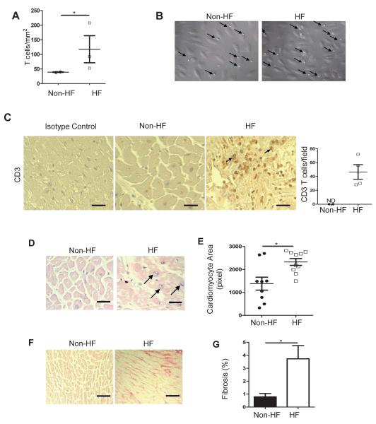
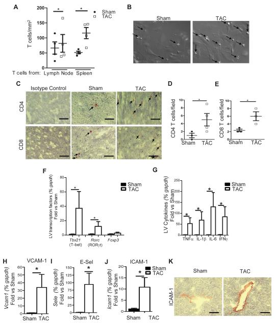
Background: Despite the emerging association between heart failure (HF) and inflammation, the role of T cells, major players in chronic inflammation, has only recently begun to be explored. Whether T-cell recruitment to the left ventricle (LV) participates in the development of HF requires further investigation to identify novel mechanisms that may serve for the design of alternative therapeutic interventions.

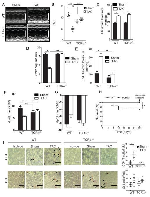
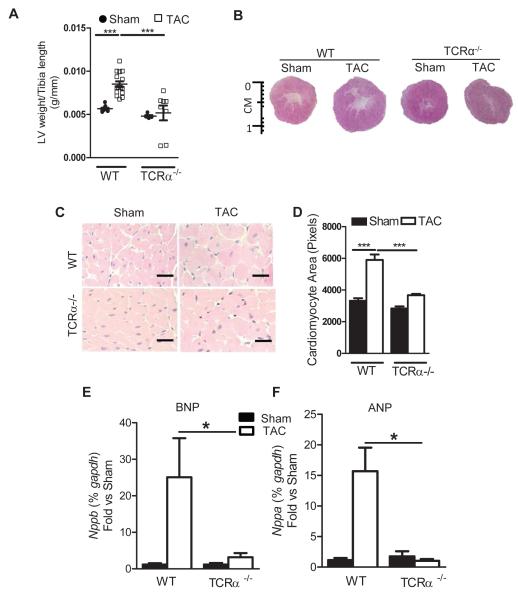
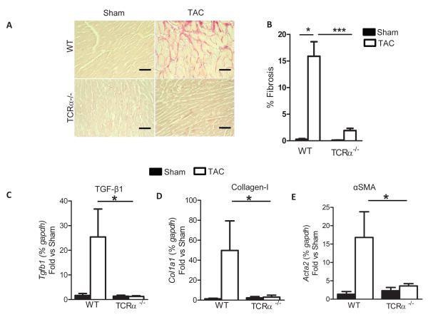
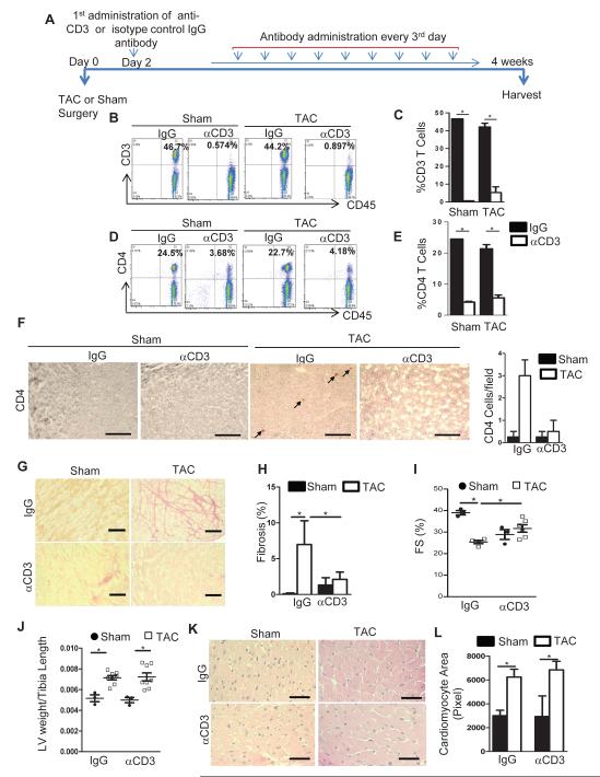
Methods and results: Real-time videomicroscopy of T cells from nonischemic HF patients or from mice with HF induced by transverse aortic constriction revealed enhanced adhesion to activated vascular endothelial cells under flow conditions in vitro compared with T cells from healthy subjects or sham mice. T cells in the mediastinal lymph nodes and the intramyocardial endothelium were both activated in response to transverse aortic constriction and the kinetics of LV T-cell infiltration was directly associated with the development of systolic dysfunction. In response to transverse aortic constriction, T cell-deficient mice (T-cell receptor, TCRα(-/-)) had preserved LV systolic and diastolic function, reduced LV fibrosis, hypertrophy and inflammation, and improved survival compared with wild-type mice. Furthermore, T-cell depletion in wild-type mice after transverse aortic constriction prevented HF.

Conclusions: T cells are major contributors to nonischemic HF. Their activation combined with the activation of the LV endothelium results in LV T-cell infiltration negatively contributing to HF progression through mechanisms involving cytokine release and induction of cardiac fibrosis and hypertrophy. Reduction of T-cell infiltration is thus identified as a novel translational target in HF.

Keywords: T lymphocytes; cell adhesion; heart failure; heart ventricles; inflammation; ventricular remodeling.


Figures

Figure 1
Figure 1. T cell activation, adhesion to activated vascular endothelial cells and infiltration into the LV in humans
(A, B) CD3+ T cells isolated from healthy volunteers or from patients with NYHA-III-IV HF were drawn in a flow chamber across TNFα (25ng/ml) activated HUVEC at a shear stress of 0.8 dynes/cm2. Data represents mean ± SD of 2 independent experiments using 2 different control and 3 different heart failure samples. Representative images from the videos are shown and arrows point at T cells arrested on the vascular endothelium (B). (C) Non-heart failure (Non-HF) and end stage HF LV tissues were obtained from NDRI and LVAD placements, respectively, and sections were stained for CD3 or isotype control. The arrows point at two representative infiltrated CD3+ T cells and the quantitative analysis is shown (right). (D, E) Representative H&E staining (D) and cardiac myocyte size quantification (E) of LV sections from non-HF and end stage HF. Arrows point to vesiculated pyramids, standard histological markers of hypertrophic myocytes. (F, G) Representative photomicrographs (F) and quantification (G) of myocardial fibrosis evaluated by picrosirius red staining of LV sections. Data represented as mean ±SEM; *p<0.05. Scale bars, 500μm (C, D and F).
Figure 2
Figure 2. TAC induces T cell adhesion to mouse heart endothelial cells in vitro and endothelial and T cell activation and recruitment to the heart in vivo
(A, B) CD4+ T cells from lymph nodes and spleen isolated from 4 weeks TAC mice or sham surgery were drawn across TNFα (100ng/ml) activated mouse heart endothelial cells at a shear stress of 0.8 dynes/cm2. Data represents mean ± SD of 4 independent adhesion experiments, each with a pool of T cells from n=3 sham and n=3-5 TAC, and 4 independent MHEC preparations. Data represents the average of 3-6 different fields of view for each condition in the different experiments (B). Representative frames from videos with arrows pointing at T cells arrested on MHEC. (C-E) LV tissue sections from 4 week sham or TAC mice were stained for CD4+ and CD8+ T cells. (C) Representative immunohistochemistry and (D, E) quantitative analysis of CD4 and CD8 staining. (F-J) RNA expression for T cell subset transcription factors (F), inflammatory cytokines (G) and adhesion molecules (H-J) in the LV of 4 week TAC WT mice. n=4-6 shams and n=6-10 TAC per group. (K) Representative immunohistochemistry staining of ICAM-1 in the LV of 4 weeks sham and TAC WT mice. Data represented as mean ±SD; *p<0.05. Scale bars, 500μm (C, K).
Figure 3
Figure 3. Time course of T cell recruitment into the LV as cardiac remodeling and systolic function develop in PO induced heart failure
WT mice were subjected to sham (n=3-11) or TAC (n= 5-16) surgery and sacrificed at the indicated times. (A) LV weight normalized to tibia length. (B) Systolic function measured by echocardiography. (C-E) Quantification of LV T cell (C), neutrophil (D) and F4/80+ macrophage (E) infiltration by FACS during the time course of TAC. (F-H) Representative FACS dot plot for total leukocytes (F) CD3 T cells (G) and CD11b+ myeloid cells (H) at 4 weeks post TAC. Numbers represent the percent positive in the indicated gate. (I) Leukocyte expression of CD3, Gr1, CD11b and F4/80 positive cells in the LV 4 weeks after sham or TAC mice. (J) Representative FACS histogram of CD62L expression in the mediastinal lymph nodes in sham and TAC mice 4 weeks post TAC surgery. Data represent mean ± SD; *p<0.05.
Figure 4
Figure 4. Preserved cardiac function and survival to PO induced heart failure in TCRα−/− mice
(A, B) Representative echocardiograms, white scale bars, 4mm (A) and quantification of fractional shortening (% FS) (B) 4 weeks after sham or TAC surgery in WT and TCRα−/− mice (n=3 Sham, and n=5 TAC). (C-G) Invasive hemodynamic measurements in WT and TCRα−/− mice measured 4 weeks after sham and TAC surgery (n=3 sham, and n=7 TAC). (H) Kaplan-Meier survival curves in WT (n=19) and TCRα−/− (n=10) mice after TAC. (I) Representative immunohistochemical staining of 4 week TAC LV sections for T cells (CD4+) and neutrophils (Gr1+). Quantification (right) of positive cells/field for n=3-5 mice. Data represent mean ±SD; *p<0.05; **p<0.005; ***p<0.0005, ns, not significant Scale bars, 500μm (I).
Figure 5
Figure 5. Absence of LV hypertrophy in response to PO induced heart failure in TCRα−/− mice
(A) LV weight, normalized to tibia length in 4 week sham or TAC mice (WT n=6-11 mice for sham and 11-16 TAC mice; TCRα−/− n=5 sham and n=8 TAC). (B) Representative pictures from LV cross sections of WT and TCRα−/− mice 4 weeks post sham or TAC surgery. (C) Representative H&E staining of LV sections from mice 4 weeks after sham or TAC surgery (D) Quantification of cardiac myocyte area (WT mice n=4 Sham and n=6 TAC; TCRα−/− mice n=4 Sham and n=7 TAC). (E, F) qPCR for hypertrophy markers BNP and ANP. n=5 sham and n=7 TAC mice. Data represented as mean ± SD. *=p<0.01, ***=p<0.0001. Scale bars, 500μm (C).
Figure 6
Figure 6. Absence of LV fibrosis in response to PO induced heart failure in TCRα−/− mice
(A) Representative photomicrographs and (B) quantification of myocardial fibrosis evaluated by picosirius red staining of LV sections from 4 week sham and TAC mice (WT: n=3-5 sham and 6 mice TAC; TCRα−/− n=4 sham and n=8 TAC). (C-E) qPCR for fibrotic markers TGFβ, Collagen I, and SMAα. Data represent mean ±SD; *p<0.05; ***p<0.005. Scale bars, 500μm (A).
Figure 7
Figure 7. T cell depletion in WT mice post TAC surgery results in preserved cardiac fibrosis and improved systolic function
(A) Diagram of experimental protocol for T cell depletion with αCD3 Ab in WT sham and TAC mice. IgG Ab was used as control. (B, D) Representative FACS dot plots (C, E) and quantification of CD3+ T cells (B,C) and CD4+ T cells (D, E) in lymph nodes of 4 week WT sham or TAC mice treated as in A. (F) Representative immunohistochemical staining of CD4+ T cells and quantification (right) of LV T cells in the indicated groups 4 week post TAC. (G) Representative photomicrographs and (H) quantification of LV fibrosis evaluated by picrosirius red staining of LV sections in the indicated groups. (I) Echocardiography analysis in Sham and TAC WT mice treated with control IgG Ab or depleted of T cells with αCD3 Ab after sham or TAC surgery. (J) LV weight, normalized to tibia length. (K) Representative H&E staining and (L) quantification of cardiac myocyte area of LV sections among the different groups. Data represent mean ±SD; *=p<0.05. Scale bars, 500μm (n=3-4 IgG and n=4-6 αCD3).

References

    1. Cowie MR, Mosterd A, Wood DA, Deckers JW, Poole-Wilson PA, Sutton GC, Grobbee DE. The epidemiology of heart failure. Eur Heart J. 1997;18:208–25. - PubMed
    1. Lloyd-Jones D, Adams R, Carnethon M, De SG, Ferguson TB, Flegal K, Ford E, Furie K, Go A, Greenlund K, Haase N, Hailpern S, Ho M, Howard V, Kissela B, Kittner S, Lackland D, Lisabeth L, Marelli A, McDermott M, Meigs J, Mozaffarian D, Nichol G, O’Donnell C, Roger V, Rosamond W, Sacco R, Sorlie P, Stafford R, Steinberger J, Thom T, Wasserthiel-Smoller S, Wong N, Wylie-Rosett J, Hong Y. Heart disease and stroke statistics--2009 update: a report from the American Heart Association Statistics Committee and Stroke Statistics Subcommittee. Circulation. 2009;11:480–6. - PubMed
    1. Ahmad T, Fiuzat M, Felker GM, O’Connor C. Novel biomarkers in chronic heart failure. Nat Rev Cardiol. 2012;9:347–59. - PubMed
    1. Jessup M, Brozena S. Heart failure. N Engl J Med. 2003;348:2007–18. - PubMed
    1. Aukrust P, Ueland T, Lien E, Bendtzen K, Muller F, Andreassen AK, Nordoy I, Aass H, Espevik T, Simonsen S, Froland SS, Gullestad L. Cytokine network in congestive heart failure secondary to ischemic or idiopathic dilated cardiomyopathy. Am J Cardiol. 1999;83:376–82. - PubMed

Publication types

MeSH terms