. 2015 Oct;63(4):917-25.
doi: 10.1016/j.jhep.2015.05.011. Epub 2015 May 27.

Monocyte-endothelial cell interactions in the regulation of vascular sprouting and liver regeneration in mouse

Affiliations

Monocyte-endothelial cell interactions in the regulation of vascular sprouting and liver regeneration in mouse

Pedro Melgar-Lesmes et al. J Hepatol. 2015 Oct.

Abstract

Background & aims: Regeneration of the hepatic mass is crucial to liver repair. Proliferation of hepatic parenchyma is intimately dependent on angiogenesis and resident macrophage-derived cytokines. However the role of circulating monocyte interactions in vascular and hepatic regeneration is not well-defined. We investigated the role of these interactions in regeneration in the presence and absence of intact monocyte adhesion.

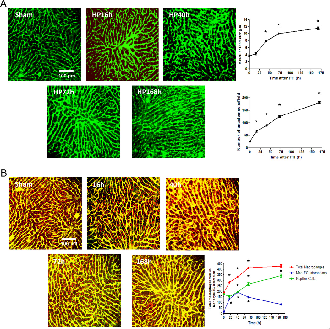
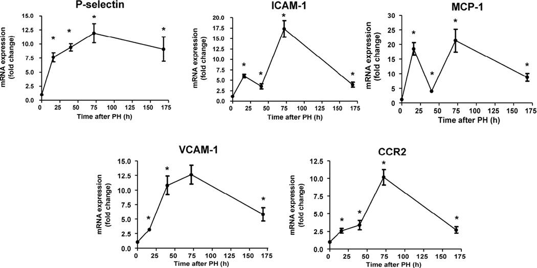
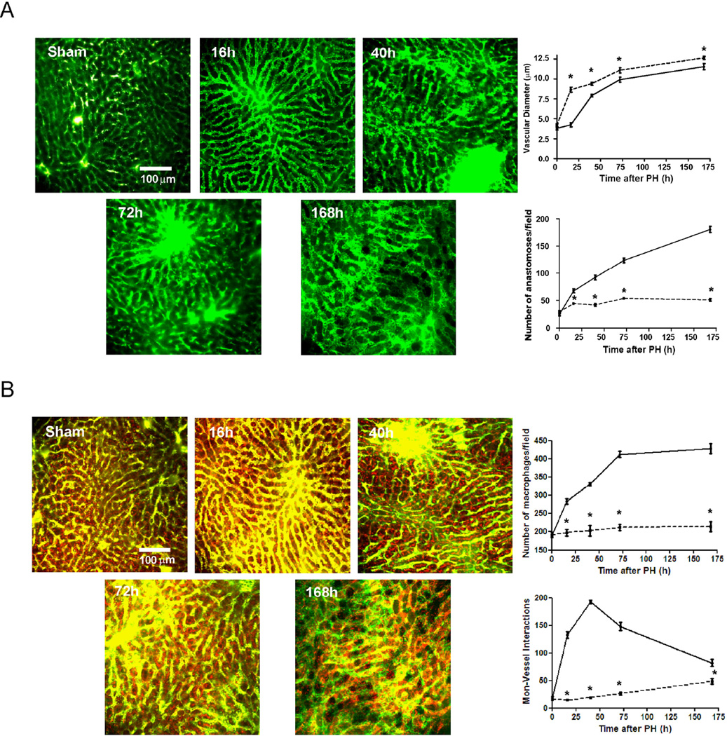
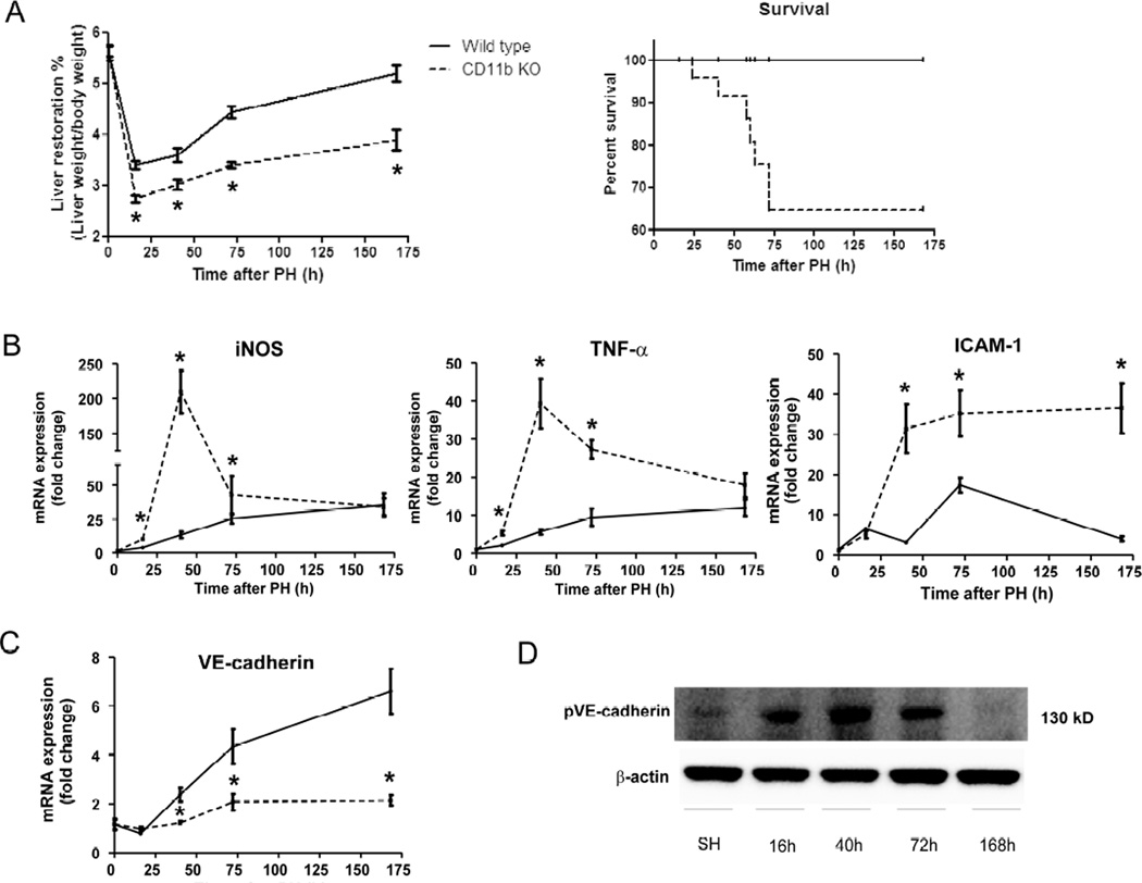
Methods: Partial hepatectomy was performed in wild-type mice and those lacking the monocyte adhesion molecule CD11b. Vascular architecture, angiogenesis and macrophage location were analyzed in the whole livers using simultaneous angiography and macrophage staining with fluorescent multiphoton microscopy. Monocyte adhesion molecule expression and sprouting-related pathways were evaluated.

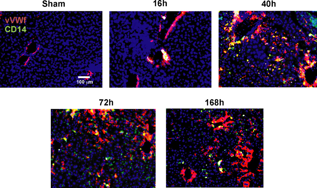
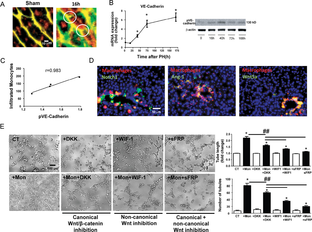
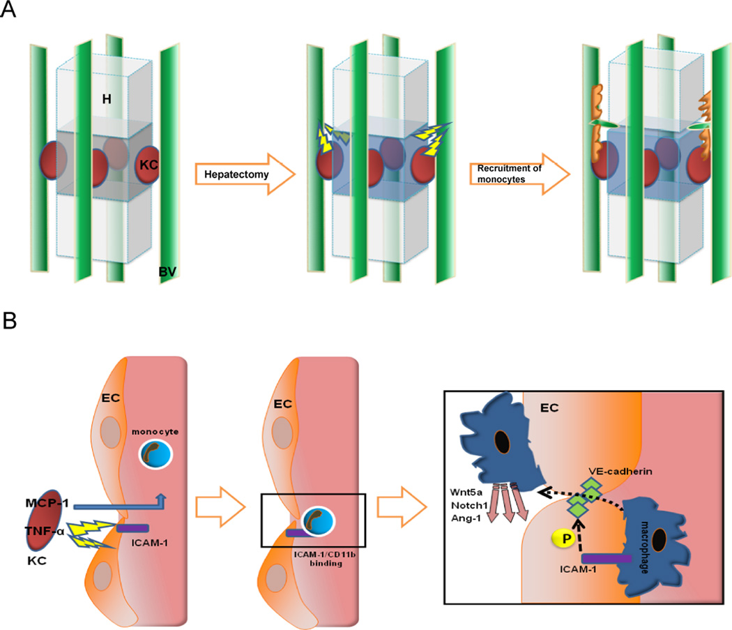
Results: Resident macrophages (Kupffer cells) did not migrate to interact with vessels whereas infiltrating monocytes were found adjacent to sprouting points. Infiltrated monocytes colocalized with Wnt5a, angiopoietin 1 and Notch-1 in contact points and commensurate with phosphorylation and disruption of VE-cadherin. Mice deficient in CD11b showed a severe reduction in angiogenesis, liver mass regeneration and survival following partial hepatectomy, and developed unstable and leaky vessels that eventually produced an aberrant hepatic vascular network and Kupffer cell distribution.

Conclusions: Direct vascular interactions of infiltrating monocytes are required for an ordered vascular growth and liver regeneration. These outcomes provide insight into hepatic repair and new strategies for hepatic regeneration.

Keywords: Angiogenesis; Hepatic repair; Immunology; Innate immune system.


Conflict of interest statement

Conflicts of interest: None declared

Figures

Figure 1
Figure 1. Monocyte/macrophage interactions with liver sinusoidal endothelial cells during liver regeneration
C57BL/6 mice underwent 70% hepatectomy and samples of liver right lobe were examined at sequential time points (0, 16, 40, 72, 168 hours post-op). Vasodilation (initiated in portal areas) and anastomoses were analyzed by angiography (vascular perfusion of FITC-dextran, MW 2×10⁶ Da) using multiphoton microscopy (A). The total number of macrophages and monocyte-vascular interactions were quantified in whole liver images obtained from mice intravenously injected with 70 kDa Texas red-dextran 2 hours before angiography with FITC-dextran and multiphoton microscopy, and sacrifice. Intravascular and extravascular monocyte-endothelium interactions and non-interacting macrophages highlighted in red were quantified in every z-stack from every field. (B); one representative Z image of liver from every time point is shown n=10 at every time point; *, p<0.05 vs. former time point.
Figure 2
Figure 2. Interactions between circulating monocytes and endothelial cells after partial hepatectomy are initiated in portal space
Representative images of liver sections identified vessels by staining for von Willebrand factor (in red) and recruited monocytes by staining for CD14 (in green). Nuclei were stained by DAPI (blue). Initial contacts of recruited monocytes (in yellow) take place in portal areas and then expand to the rest of liver sinusoid as regeneration progresses peaking at 40h post-hepatectomy.
Figure 3
Figure 3. Infiltrated macrophages stimulate vascular sprouting in contact points with vessels
Amplification of multiphoton images (vessels in green and yellow, macrophages in red), visualized vascular buds (white circles) surrounded by spread recruited macrophages as early as 16 hours after hepatectomy, none of which were identifiable in comparison to sham samples (A) mRNA levels of VE-cadherin (indicator of vascular proliferation) after hepatectomy showed that angiogenesis begins after 16 hours and progressively increases until 168 hours (B) In contrast phosphorylation of VE-cadherin (Western blot) and therefore disruption of endothelial junctions to allow the sprouting process increases from 16 to 40 hours post-op and then decreases until the end of liver regeneration (B). The pattern of phosphorylation of VE-cadherin evaluated by Western Blot shows excellent correlation with the number of infiltrated monocytes during liver regeneration (C) Hepatic staining of these recruited macrophages (CD14, in red) enhanced the local expression of sprouting-related factors Wnt5a, Notch1 and Ang-1 (in green) in contact points of vessels (yellow) in portal areas as early as 16 hours post-op (D). Tube formation assay of HUVEC inflamed by TNF-α (5 ng/mL) in the presence or absence (Control, CT) of THP-1 monocytes (Mon) or Wnt inhibitors (DKK for canonical Wnt pathway; WIF-1 for non-canonical Wnt pathway; sFRP for both Wnt pathways). Representative phase contrast images and quantification of tube length and number of tubules are shown (E). n=10 at every time point; *, p<0.05 vs. former time point; ##, p<0.01.
Figure 4
Figure 4. Sequential gene activation of ICAM-1 and MCP-1 during liver regeneration
Real-time PCR analysis of gene expression of vascular adhesion molecules (P-selectin, ICAM-1, VCAM-1 and CCR2) and the monocyte chemotactic MCP-1 showed an increasing up-regulation of P-selectin, VCAM-1 and CCR2 throughout the whole process of liver regeneration. In contrast two sequential activations of gene expression of ICAM-1 and MCP-1 were found at 16 and 72 hours post-op and a down-regulation of their transcripts at 40h thus coinciding with the peak of monocyte-EC interactions; n=10 at every time point; *, p<0.05 vs. previous time point.
Figure 5
Figure 5. Gene suppression of CD11b disrupts vascular growth, promotes a higher vasodilation and permeability and alters KC distribution after partial hepatectomy
Two groups of C57BL/6 mice (wild-type and CD11b KO) underwent 70% hepatectomy. The nature and form of the vascular network formed in the right lobe of the liver were examined sequentially (0, 16, 40, 72, 168 hours post-op) by angiography using multiphoton microscopy. Vasodilatation was present in CD11b KO mice (dashed lines) just after hepatectomy and greater than observed in wild-type mice (solid lines) during liver regeneration, but vascular growth was drastically reduced and vascular leakage visualized in CD11b KO mice as compared with wild-type mice (A). The total number of macrophages increased minimally after hepatectomy in CD11b KO mice as compared with wild-type mice. KCs migrated to interact with blood vessels after 72 hours post-op replacing the absent monocyte-ECs interactions in CD11b KO mice. (B); One representative Z image of liver from every time point in CD11b KO mice is shown; the mean values ± standard error for the groups are indicated for wild-type (solid lines) and CD11b KO (dashed lines); n=10 at every time point; *, p<0.05 vs. wild-type mice.
Figure 6
Figure 6. Mice lacking CD11b show a reduction in liver regeneration and survival after hepatectomy commensurate with up-regulation of iNOS and TNF-α
Liver samples from C57BL/6 mice subjected to 70% hepatectomy demonstrated significantly reduced hepatic mass regeneration in CD11b KO mice and as a consequence, a drop in survival in comparison to wiild-type mice (A). The absence of interactions between CD11b and ICAM-1 in blood vessels of KO mice promoted significant up-regulation of iNOS, TNF-α and ICAM-1 peaking at 40 and 72 hours post-op as compared with wild-type mice (B). Moreover mice deficient in CD11b showed a drastic reduction of VE-cadherin gene expression assessed by Real-time PCR (C) in comparison to wild-type mice and in concordance with the results showing a decrease of angiogenesis in CD11b KO mice. The up-regulation pattern of TNF-α was associated with enhanced levels of phosphorylation of VE-cadherin in CD11b KO mice peaking at 40 hours after hepatectomy (D); n=10 at every time point; *, p<0.05 vs. wild-type mice.
Figure 7
Figure 7. Proposed model of the role of circulating monocytes in liver regeneration
Tridimensional schematic structure of KC (in red), hepatocytes and blood vessels (in green) following the pattern obtained by multiphoton microscopy indicates that signals from injured liver stimulate the release of chemotactics and the induction of monocyte adhesion molecules on the endothelium. These signals recruit monocytes to selected areas, driving the sprouting and angiogenic process (A). KCs deliver MCP-1 to blood stream and TNF-α towards closer ECs to promote the expression of ICAM-1 which bind attracted monocytes which will phosphorylate interendothelial VE-cadherin to allow them to migrate throughout the vessel and locally deliver sprouting factors (B).

References

    1. Fausto N, Campbell JS, Riehle KJ. Liver regeneration. Hepatology. 2006;43:S45–S53. - PubMed
    1. Vollmar B, Menger MD. The hepatic microcirculation: mechanistic contributions and therapeutic targets in liver injury and repair. Physiol Rev. 2009;89:1269–1339. - PubMed
    1. Drixler TA, Vogten MJ, Ritchie ED, van Vroonhoven TJ, Gebbink MF, Voest EE, et al. Liver regeneration is an angiogenesis- associated phenomenon. Ann Surg. 2002;236:703–711. - PMC - PubMed
    1. Diehl AM. Neighborhood watch orchestrates liver regeneration. Nat Med. 2012;18:497–409. - PubMed
    1. Viebahn CS, Benseler V, Holz LE, Elsegood CL, Vo M, Bertolino P, et al. Invading macrophages play a major role in the liver progenitor cell response to chronic liver injury. J Hepatol. 2010;53:500–507. - PubMed

Publication types

MeSH terms