. 2015 Jun 2;65(21):2314-27.
doi: 10.1016/j.jacc.2015.03.549.

Reducing In-Stent Restenosis: Therapeutic Manipulation of miRNA in Vascular Remodeling and Inflammation

Affiliations

Reducing In-Stent Restenosis: Therapeutic Manipulation of miRNA in Vascular Remodeling and Inflammation

Robert A McDonald et al. J Am Coll Cardiol. .

Abstract

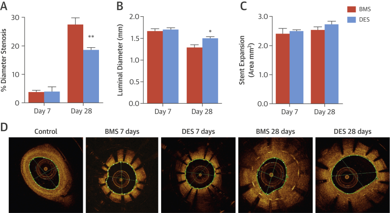
Background: Drug-eluting stents reduce the incidence of in-stent restenosis, but they result in delayed arterial healing and are associated with a chronic inflammatory response and hypersensitivity reactions. Identifying novel interventions to enhance wound healing and reduce the inflammatory response may improve long-term clinical outcomes. Micro-ribonucleic acids (miRNAs) are noncoding small ribonucleic acids that play a prominent role in the initiation and resolution of inflammation after vascular injury.

Objectives: This study sought to identify miRNA regulation and function after implantation of bare-metal and drug-eluting stents.

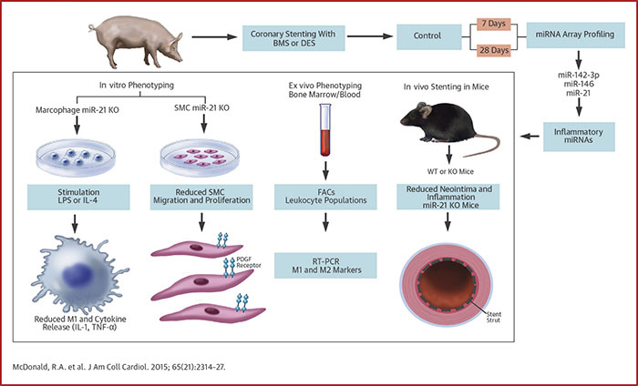
Methods: Pig, mouse, and in vitro models were used to investigate the role of miRNA in in-stent restenosis.

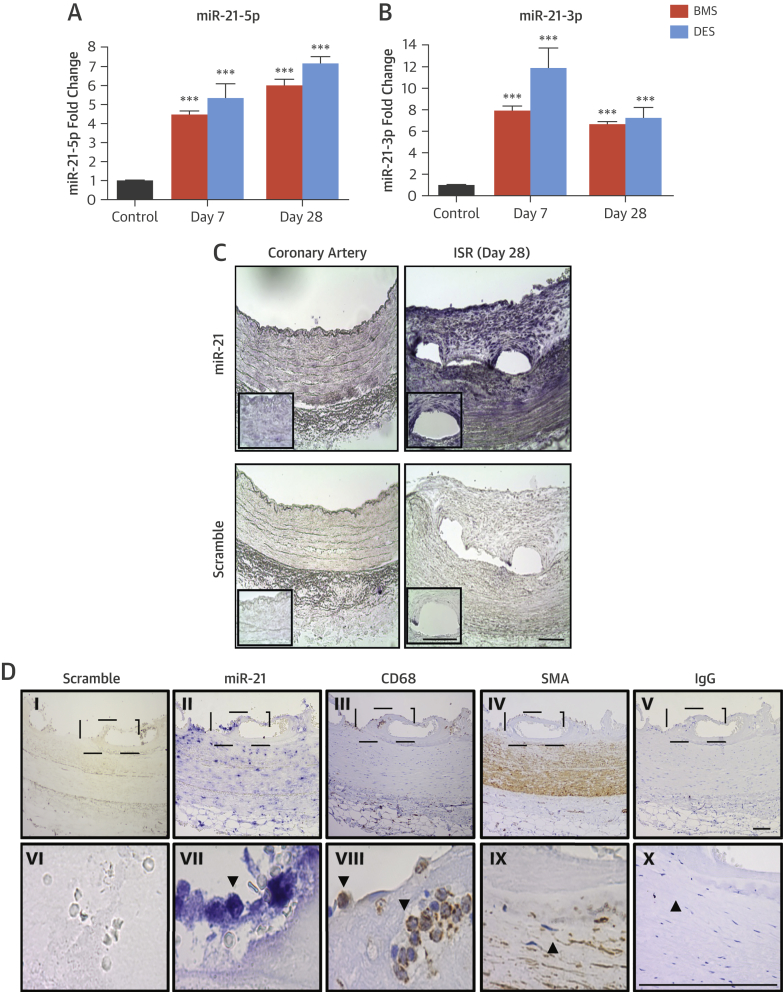
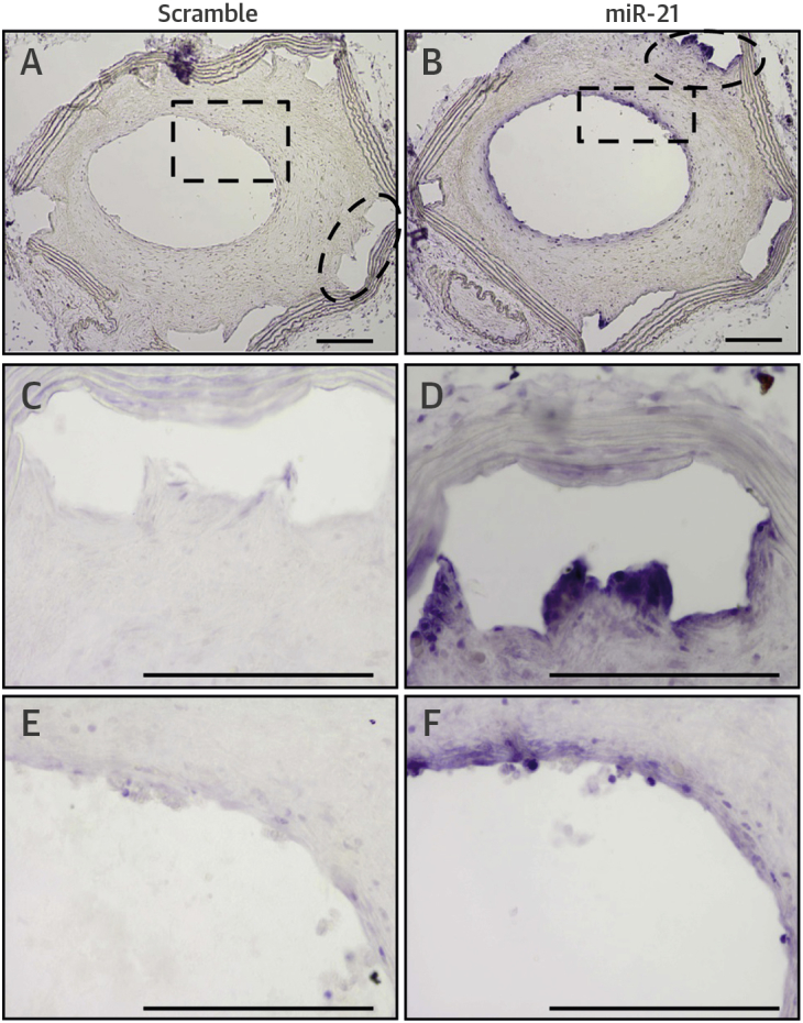
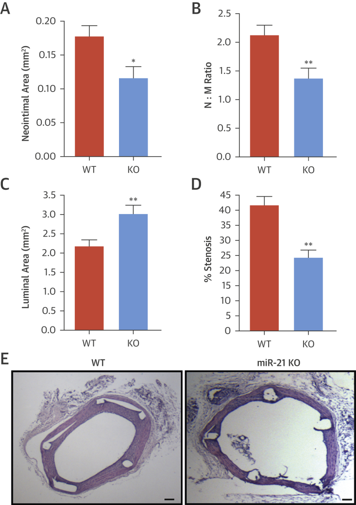
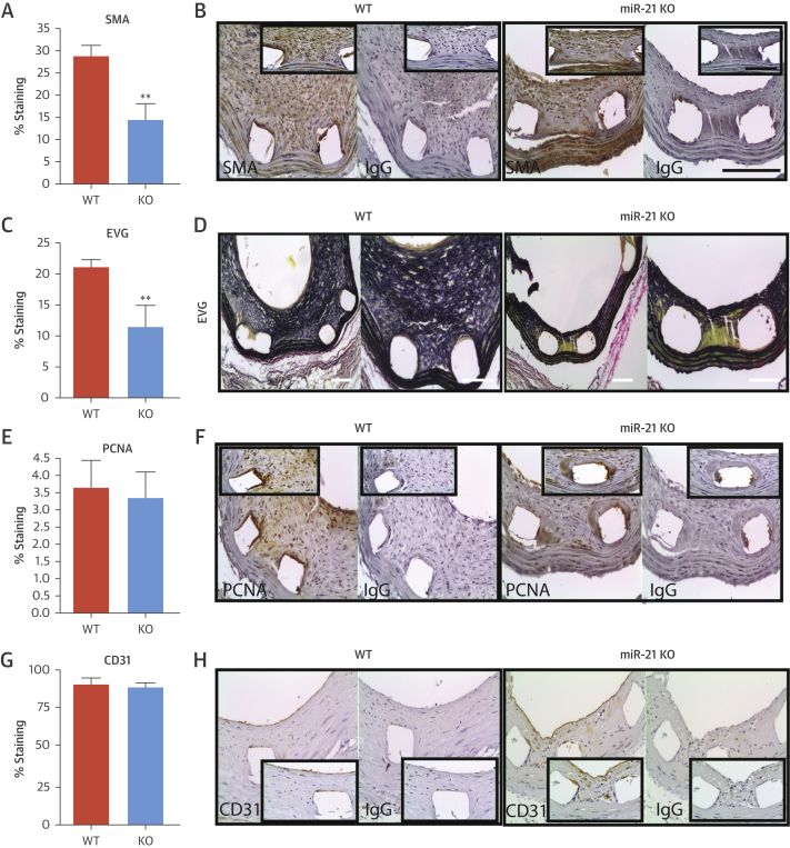
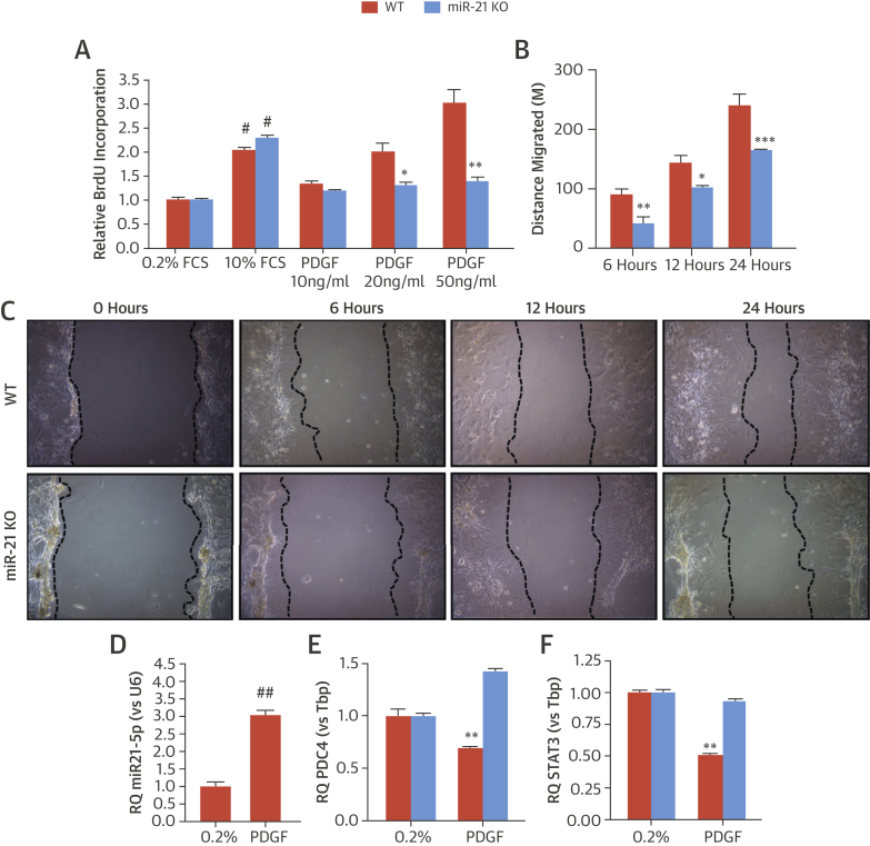
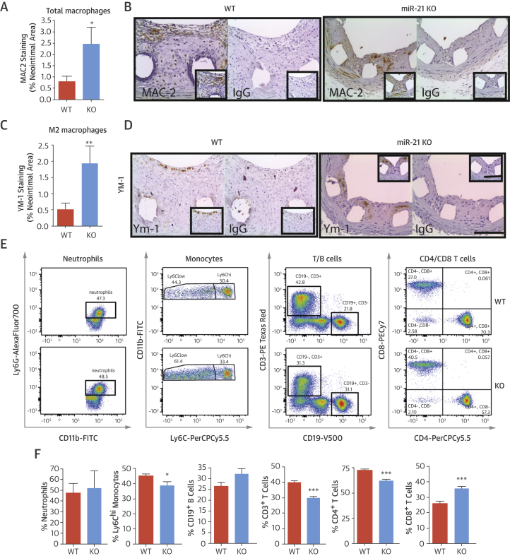
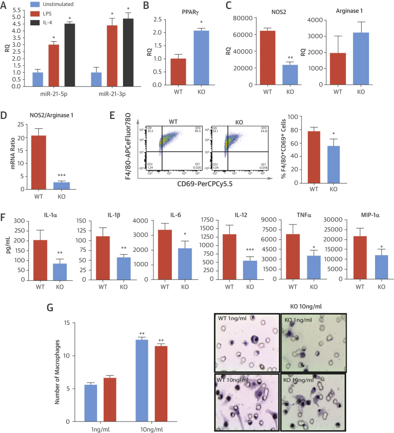
Results: We documented a subset of inflammatory miRNAs activated after stenting in pigs, including the miR-21 stem loop miRNAs. Genetic ablation of the miR-21 stem loop attenuated neointimal formation in mice post-stenting. This occurred via enhanced levels of anti-inflammatory M2 macrophages coupled with an impaired sensitivity of smooth muscle cells to respond to vascular activation.

Conclusions: MiR-21 plays a prominent role in promoting vascular inflammation and remodeling after stent injury. MiRNA-mediated modulation of the inflammatory response post-stenting may have therapeutic potential to accelerate wound healing and enhance the clinical efficacy of stenting.

Keywords: late stent thrombosis; miRNA stem loop; neointima; smooth muscle cell.


Figures

Figure 1
Figure 1
ISR Assessment: Porcine Stent Model Porcine coronary arteries were stented with bare-metal stents (BMS) or Biolimus A9 drug-eluting stents (DES). Measurements of in-stent restenosis (ISR), including (A) percent diameter stenosis, (B) luminal diameter, and (C) stent expansion, were analyzed at 7 and 28 days post-stent placement. (D) Representative optical coherence tomography images show control (unstented) arteries and arteries stented with BMS or DES for a period of 7 or 28 days. *p < 0.05; **p < 0.01 versus BMS day 28 (1-way analysis of variance).
Figure 2
Figure 2
Regulation of miR-21-5p and -3p During ISR Relative-fold change in (A) miR-21-5p and (B) miR-21-3p is expressed in control arteries or arteries stented with a BMS or DES for 7 or 28 days. ***p < 0.001 versus control arteries (1-way analysis of variance). Data are normalized to expression of U6. (C) In situ hybridization for miR-21 and scrambled control in unstented porcine coronary arteries and in BMS-stented vessels with ISR at day 28 (representative images, n = 3). Areas under enhanced magnification correspond to the regions highlighted by the hatched rectangles. Scale bar = 100 μm. (D) In situ hybridization for miR-21 and scrambled control in stented human coronary arteries and immunohistochemistry for smooth muscle actin (SMA) and cluster of differentiation (CD) 68 for smooth muscle cells and macrophages, respectively. Panels VI through X are higher magnification images of the hatch regions in panels I through V. Scale bar = 100 μm. IgG = immunoglobulin G; other abbreviations as in Figure 1.
Figure 3
Figure 3
In Situ Hybridization for miR-21 Scramble probe at (A) low magnification and (C and E) areas under enhanced magnification corresponding to regions highlighted by the hatched areas are seen in murine stented grafts 28 days post-implantation, as are (B) MiR-21 probe and (D and F) areas under enhanced magnification corresponding to regions highlighted by the hatched areas. Scale bar = 100 μm.
Figure 4
Figure 4
Effect of miR-21 Ablation on Neointimal Formation and ISR Morphometric analysis of stented aortic grafts of wild-type (WT) and miR-21 knockout (KO) mice 28 days after stent placement show (A) neointimal area, (B) neointimal/media (N/M) ratio, (C) luminal area, and (D) percent stenosis. *p < 0.05; **p < 0.01 versus WT mice. (E) Representative hematoxylin and eosin–stained sections from WT and miR-21 KO mice are seen 28 days post-stenting. Scale bar = 200 μm. Abbreviations as in Figure 1.
Figure 5
Figure 5
Cellular Analysis of Murine Lesions (A and B) The cellular composition of the neointimal lesions was quantified in WT and miR-21 KO mice at 28 days post-stenting, showing quantification of: the percentage of SMA-positive cells and representative image of the immunohistochemistry; (C and D) elastin Van Gieson (EVG) staining and representative images; (E and F) percentage of cells staining positive for proliferating cell nuclear antigen (PCNA); and (G and H) percentage of cell staining positive for CD31 within the circumference of the lumen and representative images. **p < 0.01 versus WT mice (Student unpaired t test). Scale bar = 100 μm. Abbreviations as in Figure 2, Figure 4.
Figure 6
Figure 6
In Vitro Analysis of miR-21 SMC Proliferation and migration of vascular smooth muscle cells (SMCs) were studied via proliferation assay by using (A) bromodeoxyuridine (BrdU) incorporation relative to 0.2% fetal calf serum (FCS) and (B) distance migrated by WT and KO aortic SMCs in response to platelet-derived growth factor (PDGF) 6, 12, and 24 h after stimulation. (C) Photographs represent miR-21 WT and KO SMC migration. (D) miR-21 expression levels are seen in SMCs stimulated with PDGF relative to quiesced cells (0.2% FCS). (E) Relative expression of programmed cell death protein-4 (PDCD4) and (F) signal transducer and activator of transcription 3 (STAT3) in miR-21 WT and KO SMC is significantly stimulated with PDGF. Relative quantitation (RQ) ± rqmax (vs. TATA-binding protein [Tbp]). (D) #p < 0.05 and ##p < 0.01 vs 0.2% FCS (Student unpaired t test) or (A, B, E, and F) *p < 0.05; **p < 0.01; ***p < 0.001 versus WT cells (2-way analysis of variance with Bonferroni post-hoc test). Abbreviations as in Figure 4.
Figure 7
Figure 7
Inflammatory Cells in Neointimal Lesions and Blood of miR-21 KO Mice (A) Quantification and (B) representative images of total galactose-specific lectin 3 (MAC2) staining (% neointimal area) are seen in sections of stented graft from WT and miR-21 KO mice 28 days post-stenting (scale bar = 100 μM). (C) Quantification and (D) representative images of total chitinase 3–like 3 (YM-1) staining (marker for M2 macrophages) (% neointimal area) are seen in sections of stented graft from WT and miR-21 KO mice at day 28. Flow cytometric assessment of circulating cells in blood of WT and miR-21 KO mice. (E) Representative fluorescence-activated cell sorting plots and (F) bar charts showing percent quantification of cells in gate potentially indicate a reduced ability to develop proinflammatory responses. Gating markers used: neutrophils (CD45+Ly6G+CD11b+), monocytes (CD45+Ly6G-Ly6C+CD11b+), B cells (CD45+CD3-CD19+), and T cells (CD45+CD3+CD4+ or CD45+CD3+CD8+). *p < 0.05; **p < 0.01; ***p < 0.001 versus WT mice (Student unpaired t test). Abbreviations as in Figure 2, Figure 4.
Figure 8
Figure 8
In Vitro Altered Inflammatory Response in miR-21–Deficient Macrophages (A) RQ of expression of miR-21-5p and miR-21-3p in WT macrophages stimulated with either lipopolysaccharide (LPS) or interleukin (IL)-4 for 20 h; data normalized to U6. Expression of peroxisome proliferator–activated receptor (PPAR)-γ messenger ribonucleic acid (mRNA) by quantitative polymerase chain reaction in (B) unstimulated (baseline) macrophages and (C) nitric oxide synthase (NOS2) and arginase 1 mRNA in LPS-stimulated macrophages from WT and miR-21 KO mice. Data are normalized to Tbp. (D) Ratio of expression of NOS2/arginase mRNA by quantitative polymerase chain reaction in LPS-activated macrophages was significantly higher in WT mice than in miR-21 KO mice. (E) Flow cytometric assessments found that the cell surface marker CD69 was higher in WT versus miR-21 KO macrophages after stimulation with LPS for 20 h. Representative fluorescence-activated cell sorting plot and bar chart showing quantification of data (% of F4/80+ cells expressing the marker). (F) Inflammatory cytokine (IL-1α, IL-1β, IL-6, IL-12, and TNF-α) and chemokine (macrophage inflammatory protein [MIP]-1α) production in LPS-activated macrophages was reduced in miR-21 KO compared with WT mice. (G) In a macrophage invasion and migration assay, the number of bone marrow–derived macrophages migrating though Matrigel-coated transwell inserts in response to monocyte chemoattractant protein (MCP)-1 was fixed at 6 h and quantified. Representative images of membranes are shown adjacent to graph. *p < 0.05; **p < 0.01; ***p < 0.001 versus WT mice (Student unpaired t test). Abbreviations as in Figure 2, Figure 4, Figure 6.
Central Illustration
Role of miRNA in ISR Although drug-eluting stents (DES) reduce the incidence of in-stent restenosis (ISR), they delay vascular healing and are associated with a chronic inflammatory response, which involves micro–ribonucleic acids (miRNAs). In pig, mouse, and in vitro models, miR-21 promotes vascular inflammation and remodeling after stenting and may be a therapeutic target to enhance wound healing after vascular injury. BMS = bare-metal stent(s); FACS = fluorescence-activated cell sorting; IL = interleukin; KO = knockout; LPS = lipopolysaccharide; PDGF = platelet-derived growth factor; RT-PCR = real-time polymerase chain reaction; SMC = smooth muscle cell; TNF = tumor necrosis factor; WT = wild type.

Comment in

References

    1. Serruys P.W., de Jaegere P., Kiemeneij F., et al. A comparison of balloon-expandable-stent implantation with balloon angioplasty in patients with coronary artery disease. Benestent Study Group. N Engl J Med. 1994;331:489–495. - PubMed
    1. De Bruyne B., Pijls N.H., Kalesan B., et al. Fractional flow reserve-guided PCI versus medical therapy in stable coronary disease. N Engl J Med. 2012;367:991–1001. - PubMed
    1. Andersen H.R., Nielsen T.T., Rasmussen K., et al. A comparison of coronary angioplasty with fibrinolytic therapy in acute myocardial infarction. N Engl J Med. 2003;349:733–742. - PubMed
    1. Serruys P.W., Kutryk M.J., Ong A.T. Coronary-artery stents. N Engl J Med. 2006;354:483–495. - PubMed
    1. Bavry A.A., Bhatt D.L. Appropriate use of drug-eluting stents: balancing the reduction in restenosis with the concern of late thrombosis. Lancet. 2008;371:2134–2143. - PubMed

Publication types