. 2015 Jul;56(7):1363-9.
doi: 10.1194/jlr.D059725. Epub 2015 May 28.

Quantitative lipopolysaccharide analysis using HPLC/MS/MS and its combination with the limulus amebocyte lysate assay

Affiliations

Quantitative lipopolysaccharide analysis using HPLC/MS/MS and its combination with the limulus amebocyte lysate assay

Jean-Paul Pais de Barros et al. J Lipid Res. 2015 Jul.

Abstract

Quantitation of plasma lipopolysaccharides (LPSs) might be used to document Gram-negative bacterial infection. In the present work, LPS-derived 3-hydroxymyristate was extracted from plasma samples with an organic solvent, separated by reversed phase HPLC, and quantitated by MS/MS. This mass assay was combined with the limulus amebocyte lysate (LAL) bioassay to monitor neutralization of LPS activity in biological samples. The described HPLC/MS/MS method is a reliable, practical, accurate, and sensitive tool to quantitate LPS. The combination of the LAL and HPLC/MS/MS analyses provided new evidence for the intrinsic capacity of plasma lipoproteins and phospholipid transfer protein to neutralize the activity of LPS. In a subset of patients with systemic inflammatory response syndrome, with documented infection but with a negative plasma LAL test, significant amounts of LPS were measured by the HPLC/MS/MS method. Patients with the highest plasma LPS concentration were more severely ill. HPLC/MS/MS is a relevant method to quantitate endotoxin in a sample, to assess the efficacy of LPS neutralization, and to evaluate the proinflammatory potential of LPS in vivo.

Keywords: diagnostic tool; human; inflammation; lipid transfer protein; lipoprotein; liquid chromatography tandem mass spectrometry; mass spectrometry; mouse; sepsis; systemic inflammatory response syndrome.


Figures

Fig. 1.
Fig. 1.
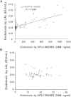
Comparison of sensitivity and accuracy of HPLC/MS/MS and GC/MS techniques for LPS quantitation in plasma samples. Mouse plasma samples containing variable amounts of LPS were obtained after intraperitoneal injection of LPS from E. coli (O55:B5) and were subjected to HCl hydrolysis and lipid extraction as described in Materials and Methods. One aliquot of the resulting preparation was analyzed by HPLC/MS/MS, and another aliquot was analyzed by GC/MS for quantitation of 3HM and calculation of plasma LPS concentration (see Materials and Methods). A: Linear regression curve built by plotting LPS values determined by GC/MS against LPS values determined by HPLC/MS/MS. B: Linear regression curve as well as the determination of the LOD and LOQ of HPLC/MS/MS and GC/MS by using the lowest values (0–40 ng/ml range) of LPS levels in mouse plasma.
Fig. 2.
Fig. 2.
Comparison of sensitivity and accuracy of HPLC/MS/MS and LAL techniques for LPS quantitation in plasma samples. Mouse plasma samples containing variable amounts of LPS were obtained after intraperitoneal injection of LPS from E. coli (O55:B5) and blood collection by retroorbital puncture. For each sample, one aliquot was directly analyzed by LAL assay, and another aliquot was subjected to HCl hydrolysis and lipid extraction prior to quantitation of 3HM by HPLC/MS/MS and calculation of plasma LPS concentration (see Materials and Methods). A: Linear regression curve built by plotting LPS values determined by LAL assay against LPS values determined by HPLC/MS/MS. B: The determination of the LOD and the LOQ of HPLC/MS/MS and LAL assay by using the lowest values (0–40 ng/ml range) of LPS levels in mouse plasma; no significant correlation between LAL and HPLC/MS/MS was observed in the low range of LPS levels (R2 = 7E–5).
Fig. 3.
Fig. 3.
Effect of incubation on the 3HM levels determined by HPLC/MS/MS in human plasma. Total human plasma was spiked with known amounts of LPS (closed diamonds, 300 ng/ml; open diamonds, 600 ng/ml; closed circles, 1,200 ng/ml; open circles, 2,400 ng/ml), and incubated at 37°C for 15 min, 30 min, 60 min, 90 min, 2 h, or 4 h. Incubated samples were analyzed by HPLC/MS/MS for quantitation of 3HM (A) and by LAL assay (B) as described in Materials and Methods.
Fig. 4.
Fig. 4.
Plasma kinetic curves of LPS levels determined by HPLC/MS/MS and LAL assay in wild-type mice. Wild-type C57BL/ mice (n = 6) were injected intraperitoneally with LPS from E. coli (O55:B5). Blood samples were drawn by retroorbital puncture before injection (time 0) and 0.5, 1, 3, 6, 24, and 48 h after injection, and plasma was obtained after centrifugation. For each plasma sample, one aliquot was analyzed by LAL assay (closed circles) and another by HPLC/MS/MS (open circles) for LPS quantitation as described in Materials and Methods. Values are expressed in ng/ml (for HPLC/MS/MS) or in endotoxin units (EU)/mL (for LAL) and are the means of 6 animals ± SEM.
Fig. 5.
Fig. 5.
Ratio of LPS activity to LPS mass concentration determined in wild-type and PLTP-deficient mice. Blood samples were drawn by retroorbital puncture before injection (time 0) and 0.5, 1.5, 3, and 6 h after injection of E. coli LPS (O55:B5, 1 mg/kg body weight). Plasma was obtained after centrifugation. LPS biological activity (LAL assay) to LPS mass concentration (direct 3-OH-myristate assay) ratios were determined over the 6 h period in wild-type (dotted line) and PLTP-deficient (PLTP−/−) mice (full line). Values are mean ± SEM of 6 animals. ** P < 0.01, significantly different from wild-type mice.

References

    1. Vincent J. L., Opal S. M., Marshall J. C., Tracey K. J. 2013. Sepsis definitions: time for change. Lancet. 381: 774–775. - PMC - PubMed
    1. Fitting C., Parlato M., Adib-Conquy M., Memain N., Philippart F., Misset B., Monchi M., Cavaillon J. M., Adrie C. 2012. DNAemia detection by multiplex PCR and biomarkers for infection in systemic inflammatory response syndrome patients. PLoS ONE. 7: e38916. - PMC - PubMed
    1. Gibot S., Béné M. C., Noel R., Massin F., Guy J., Cravoisy A., Barraud D., De Carvalho Bittencourt M., Quenot J. P., Bollaert P. E., et al. 2012. Combination biomarkers to diagnose sepsis in the critically ill patient. Am. J. Respir. Crit. Care Med. 186: 65–71. - PubMed
    1. Quenot J. P., Binquet C., Kara F., Martinet O., Ganster G. F., Navellou J. C., Castelain V., Barraud D., Cousson J., Louis G., et al. 2013. The epidemiology of septic shock in French intensive care units: the prospective multicenter cohort EPISS study. Crit. Care. 17: R65. - PMC - PubMed
    1. Beutler B., Rietschel E. T. 2003. Innate immune sensing and its roots: the story of endotoxin. Nat. Rev. Immunol. 3: 169–176. - PubMed

Publication types

LinkOut - more resources