. 2015 Jun 15;142(12):2163-72.
doi: 10.1242/dev.117341. Epub 2015 May 28.

Dnmt1 is essential to maintain progenitors in the perinatal intestinal epithelium

Affiliations

Dnmt1 is essential to maintain progenitors in the perinatal intestinal epithelium

Ellen N Elliott et al. Development. .

Abstract

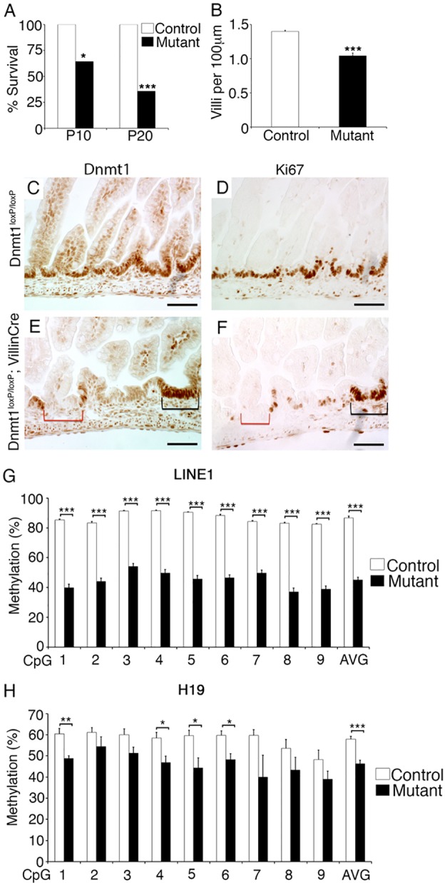
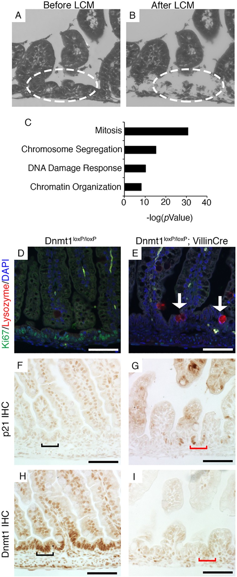
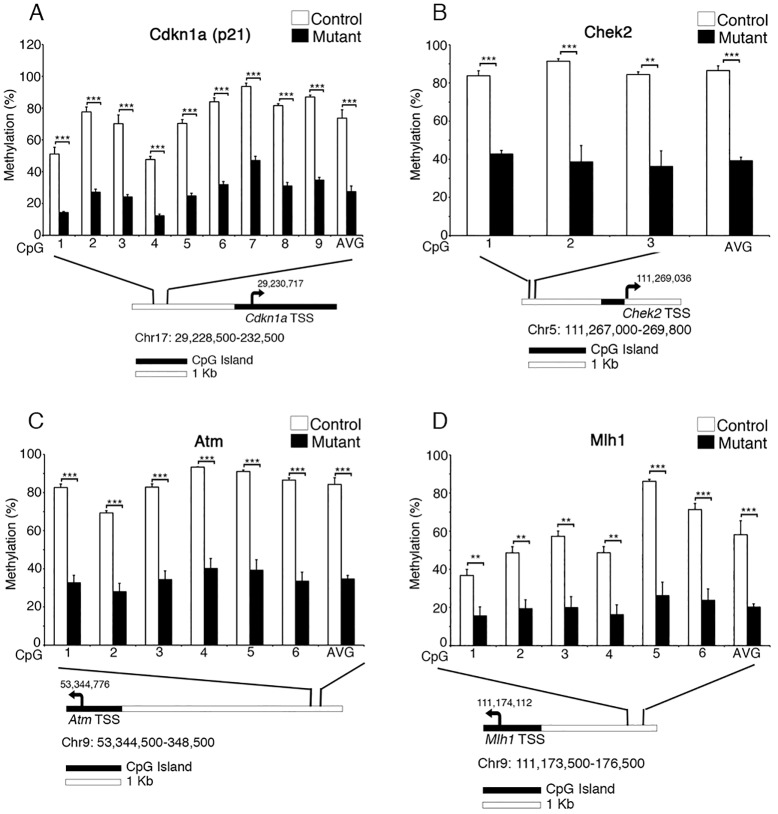
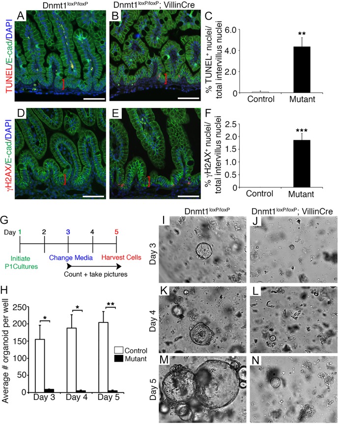
The DNA methyltransferase Dnmt1 maintains DNA methylation patterns and genomic stability in several in vitro cell systems. Ablation of Dnmt1 in mouse embryos causes death at the post-gastrulation stage; however, the functions of Dnmt1 and DNA methylation in organogenesis remain unclear. Here, we report that Dnmt1 is crucial during perinatal intestinal development. Loss of Dnmt1 in intervillus progenitor cells causes global hypomethylation, DNA damage, premature differentiation, apoptosis and, consequently, loss of nascent villi. We further confirm the crucial role of Dnmt1 during crypt development using the in vitro organoid culture system, and illustrate a clear differential requirement for Dnmt1 in immature versus mature organoids. These results demonstrate an essential role for Dnmt1 in maintaining genomic stability during intestinal development and the establishment of intestinal crypts.

Keywords: DNA damage response; DNA methylation; Intestinal epithelial development.


Figures

Fig. 1.
Fig. 1.
Ablation of Dnmt1 in the developing intestinal epithelium causes reduced proliferation and DNA hypomethylation. (A) Only 35% of Dnmt1loxP/loxP; VillinCre mouse mutants survive to P20 (n=14). (B) P0 mutants have significantly fewer villi than littermate controls (n=4). (C-F) Dnmt1 (C,E) and Ki67 (D,F) protein immunohistochemistry. P0 mutants display mosaic Dnmt1 ablation compared with controls (C,E). Absence of Dnmt1 (E, red bracket) corresponds to loss of proliferation in mutant progenitors as assessed by Ki67 staining of an adjacent section (F, red bracket). Dnmt1+ progenitors show similar proliferation in mutants (E,F, black brackets) compared with controls (D). (G) LINE1 repeat DNA methylation levels as assessed by bisulfite sequencing (n=4). Decreased LINE1 methylation indicates global demethylation in Dnmt1 mutant progenitor cells as compared with controls. (H) H19 ICR DNA methylation levels are decreased in Dnmt1 mutant progenitor cells relative to controls (n=4). Data are represented as mean±s.e.m. *P<0.05, **P<0.01, ***P<0.001, two-tailed Student's t-test. Scale bars: 50 μm.
Fig. 2.
Fig. 2.
RNA-Seq analysis reveals decreased expression of crucial cell-cycle regulators in Dnmt1-deficient progenitor cells. (A,B) Collection of intervillus regions (encircled) before (A) and after (B) LCM. (C) DAVID GO analysis of genes with transcripts that are downregulated in P0 Dnmt1-deficient epithelial cells as determined by RNA-Seq. (D,E) Co-staining for Ki67 (green) and lysozyme (red) in control and Dnmt1 mutant intestine. A fraction of non-replicating Dnmt1 cells exhibits increased lysozyme protein levels (E, arrows), a marker for Paneth cell differentiation, relative to controls (D). (F,G) Immunohistochemistry (IHC) showing that in controls p21 protein is confined to differentiated cells in the villus; in Dnmt1 mutants, expression is strikingly increased in intervillus regions (black bracket in F versus red bracket in G). (H,I) Dnmt1 immunohistochemistry on serial sections confirms Dnmt1 ablation in cells indicated by red brackets in G,I. Scale bars: 50 μm.
Fig. 3.
Fig. 3.
Key DNA damage response genes are significantly demethylated in Dnmt1-ablated perinatal intestinal epithelial progenitors. Targeted bisulfite sequencing analysis of DNA damage response genes demonstrates significant demethylation upstream of p21 (Cdkn1a) (A), Chek2 (B), Atm (C) and Mlh1 (D) in P0 Dnmt1loxP/loxP; VillinCre mutants as compared with littermate controls. Beneath each bar chart is a diagram indicating the position of the transcription start site (TSS, arrow) and CpG islands relative to the regions sequenced. Each region is ∼2-4 kb upstream of the TSS. **P<0.01, ***P<0.001, two-tailed Student's t-test.
Fig. 4.
Fig. 4.
Ablation of Dnmt1 in the immature intestine results in loss of progenitor cells via apoptosis in vivo and in vitro. (A,B) TUNEL staining (red) demonstrates increased levels of apoptosis in P0 Dnmt1 mutant progenitor cells (A) as compared with the control (B). (C) TUNEL+ intervillus nuclei within 20 µm of the epithelial base (red brackets in A,B) were quantified in Dnmt1 mutants and littermate controls (n≥5). (D,E) Dnmt1 mutant progenitors (E) have increased DNA double-strand breaks compared with controls (D), as determined by γH2AX staining (red). (F) Quantification of γH2AX+ cell number as performed for TUNEL+ cells (n=4) within 20 µm of the epithelial base (red brackets in D,E). (A,B,D,E) E-cadherin immunostaining (green) to outline the epithelium and DAPI staining (blue) of nuclei. (G) Experimental scheme for P1 Dnmt1loxP/loxP (n=3) and Dnmt1loxP/loxP; VillinCre (n=2) intestinal organoid cultures. (H) Average number of organoids per well during the timecourse of the experiment. Organoids were grown in triplicate for each biological replicate. (I-N) Live phase-contrast imaging of Dnmt1loxP/loxP control and Dnmt1loxP/loxP; VillinCre mutant organoids on each day of culture. Surviving mutant organoids on day 5 persisted as very small cysts (N), whereas controls were considerably larger and exhibited budding activity (M). Phase-contrast images were all captured at 10× magnification. Error bars indicate s.e.m. *P<0.05, **P<0.01, ***P<0.001, two-tailed (C,F) or one-tailed (H) Student's t-test. Scale bars: 50 μm.

References

    1. Ahuja N., Mohan A. L., Li Q., Stolker J. M., Herman J. G., Hamilton S. R., Baylin S. B. and Issa J. P. (1997). Association between CpG island methylation and microsatellite instability in colorectal cancer. Cancer Res. 57, 3370-3374. - PubMed
    1. Allan L. A., Duhig T., Read M. and Fried M. (2000). The p21(WAF1/CIP1) promoter is methylated in Rat-1 cells: stable restoration of p53-dependent p21(WAF1/CIP1) expression after transfection of a genomic clone containing the p21(WAF1/CIP1) gene. Mol. Cell. Biol. 20, 1291-1298. 10.1128/MCB.20.4.1291-1298.2000 - DOI - PMC - PubMed
    1. Al-Nafussi A. I. and Wright N. A. (1982). Cell kinetics in the mouse small intestine during immediate postnatal life. Virchows Arch. B Cell Pathol. Incl. Mol. Pathol. 40, 51-62. 10.1007/BF02932850 - DOI - PubMed
    1. Ashburner M., Ball C. A., Blake J. A., Botstein D., Butler H., Cherry J. M., Davis A. P., Dolinski K., Dwight S. S., Eppig J. T. et al. (2000). Gene ontology: tool for the unification of biology. The Gene Ontology Consortium. Nat. Genet. 25, 25-29. 10.1038/75556 - DOI - PMC - PubMed
    1. Baubonis W. and Sauer B. (1993). Genomic targeting with purified Cre recombinase. Nucleic Acids Res. 21, 2025-2029. 10.1093/nar/21.9.2025 - DOI - PMC - PubMed

Publication types

MeSH terms

Substances

Associated data