. 2015 May 29;348(6238):1024-7.
doi: 10.1126/science.aaa4559.

Cell adhesion. Mechanical strain induces E-cadherin-dependent Yap1 and β-catenin activation to drive cell cycle entry

Affiliations

Cell adhesion. Mechanical strain induces E-cadherin-dependent Yap1 and β-catenin activation to drive cell cycle entry

Blair W Benham-Pyle et al. Science. .

Abstract

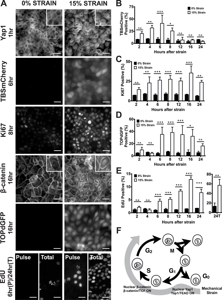
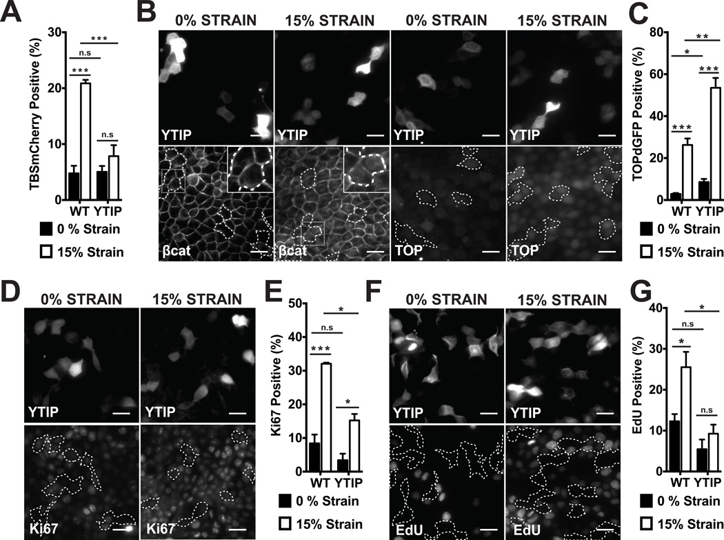
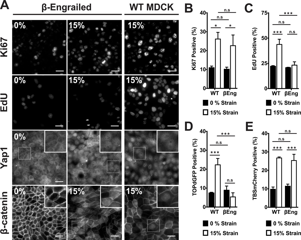
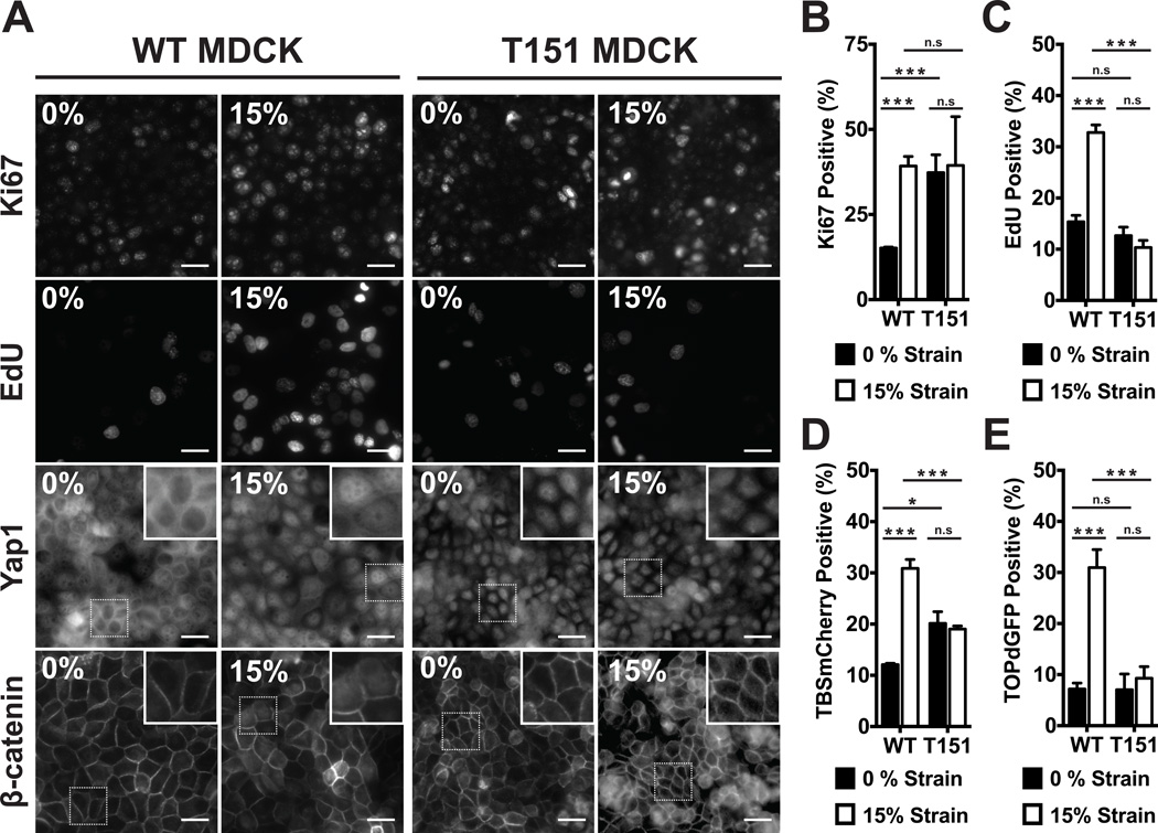
Mechanical strain regulates the development, organization, and function of multicellular tissues, but mechanisms linking mechanical strain and cell-cell junction proteins to cellular responses are poorly understood. Here, we showed that mechanical strain applied to quiescent epithelial cells induced rapid cell cycle reentry, mediated by independent nuclear accumulation and transcriptional activity of first Yap1 and then β-catenin. Inhibition of Yap1- and β-catenin-mediated transcription blocked cell cycle reentry and progression through G1 into S phase, respectively. Maintenance of quiescence, Yap1 nuclear exclusion, and β-catenin transcriptional responses to mechanical strain required E-cadherin extracellular engagement. Thus, activation of Yap1 and β-catenin may represent a master regulator of mechanical strain-induced cell proliferation, and cadherins provide signaling centers required for cellular responses to externally applied force.


Figures

Figure 1
Figure 1
Mechanical strain induces cell cycle re-entry and sequential activation of Yap1 and β-catenin. (A) Distribution of Yap1 1hr after no strain or 15% strain, TBSmCherry after 6hrs, Ki67 after 8hrs, β-catenin after 16hrs, TOPdGFP after 16hrs, and EdU incorporation after a 1hr pulse (‘Pulse’) before fixation at 6hrs, or total EdU incorporation over 24hrs (‘Total’). Insets show higher magnification of the region outlined by a dotted line. (B–E) Levels of TBSmCherry (B), Ki67 (C), TOPdGFP (D) and EdU (E) in MDCK monolayers 2–24hrs after mechanical strain; for methods used for quantification, see Supplementary Information. (F) Summary of cell cycle responses to mechanical strain. Scale bars: 25 µm. All quantifications were from at least 3 independent experiments with 2 replicate monolayers per experiment (Table S1). Quantifications were mean +/− SEM; unpaired t-test p values < 0.05 (*), <0.01(**) and <0.001 (***).
Figure 2
Figure 2
Yap1-mediated transcription is required for cell cycle re-entry after mechanical strain, but not for β-catenin nuclear accumulation or transcriptional activity. (A) % TBSmCherry-positive cells in MDCK monolayers 4hrs after no strain or 15% strain in control (WT) and YTIP-GFP expressing cells; (B) β-catenin and TOPdGFP distributions in YTIP-RFP expressing cells and TOPdGFP quantification (C) after 8 hours; (D) Ki67 distribution and quantification (E) after 8hrs; EdU incorporation (F) and quantification (G) after 24hrs in WT or YTIP-expressing cells after no strain or 15% strain. YTIP-expressing cells outlined with white dashed lines; insets show higher magnification of the region outlined by a dotted line. Scale bars: 25 µm. All quantifications (see Supplementary Information) were from at least 3 independent experiments with 2 replicate monolayers per experiment (Table S1). Quantifications were mean +/− SEM; unpaired t-test p values < 0.05 (*), p values < 0.01(**), p values <0.001 (***).
Figure 3
Figure 3
β-catenin transcriptional activity was required for cell cycle progression through G1 into S phase after mechanical strain. (A) Distributions of Yap1 4hrs after no strain or 15% strain, Ki67 and β-catenin after 8hrs, and EdU incorporation after 24hrs in control (WT) and βEng-expressing MDCK monolayers. Insets show higher magnification of the region outlined by a dotted line. Quantification of Ki67 (B) and TOPdGFP (D) 8hrs after no strain or 15% strain, EdU (C) after 24hrs, and TBSmCherry (E) after 4hrs in control (WT) and βEng MDCK cells. Scale bars: 25 µm. All quantifications (see Supplementary Information) were from at least 3 independent experiments with 2 replicate monolayers per experiment (Table S1). Quantifications were mean +/− SEM; unpaired t-test p values < 0.05 (*), p values < 0.01(**), p values <0.001 (***).
Figure 4
Figure 4
E-cadherin extracellular domain interactions were required for quiescence at high density, Yap1 nuclear exclusion, and β-catenin activation following strain. (A) Distribution of Yap1 after 4hrs, Ki67 and β-catenin after 8hrs, and EdU incorporation after 24hrs in control (WT) and T151 MDCK monolayers after no strain or 15% strain. Insets show higher magnification of the region outlined by a dotted line. Quantification of Ki67 (B) and TOPdGFP (E) after 8hrs, EdU (C) after 24hrs, and TBSmCherry (D) after 4hrs in control (WT) and T151 MDCK cell monolayers after no strain or 15% strain. Scale bars: 25 µm. All quantifications (see Supplementary Information) were from least 3 independent experiments with 2 replicate monolayers per experiment (Table S1). Quantifications were mean +/− SEM; unpaired t-test p values < 0.05 (*), p values <0.001 (***).

Comment in

References

    1. Chen CS, Tan J, Tien J. Mechanotransduction at cell-matrix and cell-cell contacts. Annu. Rev. Biomed. Eng. 2004;6:275–302. - PubMed
    1. Streichan SJ, Hoerner CR, Schneidt T, Holzer D, Hufnagel L. Spatial constraints control cell proliferation in tissues. Proceedings of the National Academy of Sciences. 2014;111:5586–5591. - PMC - PubMed
    1. Gilbert PM, et al. Substrate Elasticity Regulates Skeletal Muscle Stem Cell Self-Renewal in Culture. Science. 2010;329:1078–1081. - PMC - PubMed
    1. Engler AJ, Sen S, Sweeney HL, Discher DE. Matrix Elasticity Directs Stem Cell Lineage Specification. Cell. 2006;126:677–689. - PubMed
    1. Dupont S, et al. Role of YAP/TAZ in mechanotransduction. Nature. 2011;474:179–183. - PubMed

Publication types

MeSH terms