. 2015 Jun 9;112(23):E3010-9.
doi: 10.1073/pnas.1505870112. Epub 2015 May 28.

Transcription factor PRDM8 is required for rod bipolar and type 2 OFF-cone bipolar cell survival and amacrine subtype identity

Affiliations

Transcription factor PRDM8 is required for rod bipolar and type 2 OFF-cone bipolar cell survival and amacrine subtype identity

Cynthia C Jung et al. Proc Natl Acad Sci U S A. .

Abstract

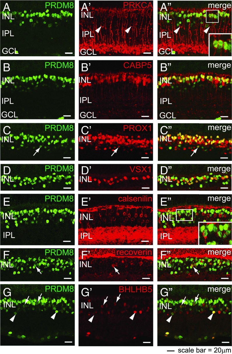
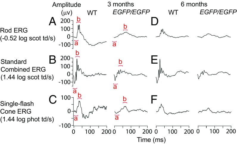
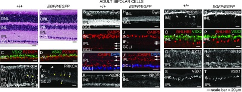
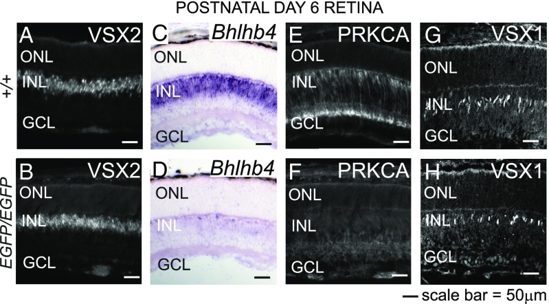
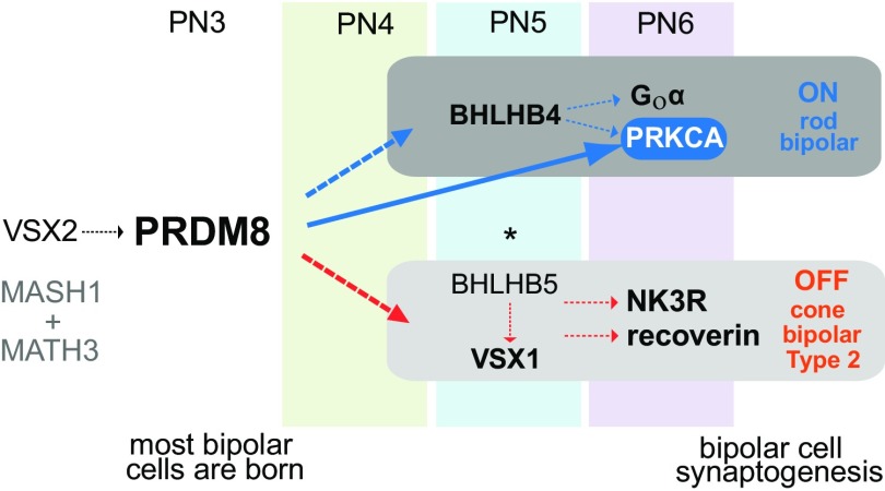
Retinal bipolar (BP) cells mediate the earliest steps in image processing in the visual system, but the genetic pathways that regulate their development and function are incompletely known. We identified PRDI-BF1 and RIZ homology domain containing 8 (PRDM8) as a highly conserved transcription factor that is abundantly expressed in mouse retina. During development and in maturity, PRDM8 is expressed strongly in BP cells and a fraction of amacrine and ganglion cells. To determine whether Prdm8 is essential to BP cell development or physiology, we targeted the gene in mice. Prdm8(EGFP/EGFP) mice showed nonprogressive b-wave deficits on electroretinograms, consistent with compromised BP cell function or circuitry resembling the incomplete form of human congenital stationary night blindness (CSNB). BP cell specification was normal in Prdm8(EGFP/EGFP) retina as determined by VSX2(+) cell numbers and retinal morphology at postnatal day 6. BP subtype differentiation was impaired, however, as indicated by absent or diminished expression of BP subtype-specific markers, including the putative PRDM8 regulatory target PKCα (Prkca) and its protein. By adulthood, rod bipolar (RB) and type 2 OFF-cone bipolar (CB) cells were nearly absent from Prdm8-null mice. Although no change was detected in total amacrine cell (AC) numbers, increased PRKCA(+) and cholinergic ACs and decreased GABAergic ACs were seen, suggesting an alteration in amacrine subtype identity. These findings establish that PRDM8 is required for RB and type 2 OFF-CB cell survival and amacrine subtype identity, and they present PRDM8 as a candidate gene for human CSNB.

Keywords: amacrine cell; bipolar cell; development; genetics; retina.


Conflict of interest statement

The authors declare no conflict of interest.

Figures

Fig. 1.
Fig. 1.
PRDM8 is expressed in differentiating neurons in all layers of the developing neural retina. Representative confocal micrographs of sections from developing WT retina were costained with antibodies to PRDM8 and cell type-specific markers. (A) PRDM8 was expressed in postmitotic BRN3B+ RGCs at E12.5. (B) ISL1 is an early marker of cholinergic ACs and RGCs. At E18.5, Prdm8 was expressed in a subset of developing ISL1+ RGCs or displaced ACs in the GCL (arrows), but ISL1 and PRDM8 were expressed in different AC populations in the nascent INL (AC[). A few faintly PRDM8+ oblong cells, with the appearance of postmitotic migrating neurons, were present in the ONBL from E18.5 to PN6 (white arrowheads in B–E). (C) At PN1, the pattern of PRDM8 expression in RGCs and a band of ACs was similar to that at E18.5. (D) Low-intensity PRDM8 expression in BP cells was first observed at PN3 (yellow arrowheads and ]BP). (E and F) At PN6, PRDM8 was expressed widely among differentiating VSX2+ BP cells, (G) in PAX6+ ACs in the INL (arrows) and PAX6+ RGCs and/or displaced ACs in the GCL (arrowheads), and (H) in BRN3+ RGCs (arrowheads). (I–L) In adult retina, PRDM8 expression was maintained in all three nuclear layers but at a very low level in PRs [outer nuclear layer (ONL)]. (J) PRDM8 was coexpressed in a subset of VSX2+ BP cells, (K) a small fraction of PAX6+ ACs in the INL (arrows) and PAX6+ RGCs and/or displaced ACs in the GCL (arrowheads), and (L) some BRN3+ RGCs. le, lens.
Fig. 2.
Fig. 2.
PRDM8 is expressed in adult BP cell subtypes. Representative confocal micrographs showing PRDM8 antibody costaining with BP subtype markers on WT retinal sections. (A–G) PRDM8 staining showed nuclear expression, which was most intense at the outer edge of the INL and of variable intensity in the rest of the INL. (A′) PRKCA stained intensely the perimeter of RB cell bodies at the outer edge of the INL, RB axons, dendrites, and a small number of ACs toward the inner edge of the INL (arrowheads). (A″) PRDM8+ nuclei were found within all PRKCA+ RB cell bodies (Inset shows an enlargement of the boxed area) but not within PRKCA+ ACs (arrowheads). (B′) CABP5 marks the nuclei and axons of types 3 and 5 CB and RB cells. (B″) PRDM8 colocalized with many CABP5+ nuclei at the upper edge of the INL but not with CABP5+ nuclei located mid-INL. (C) The nuclear marker PROX1 stains RB cells, horizontal cells, undefined CB subtypes, and AII ACs (arrow). (C″) PRDM8 colocalized with many PROX1+ BP cells in the outer INL but only a rare subset of PROX1+ ACs in the basal INL (arrow) (Fig. S1 G–H). (DD″) PRDM8 was expressed in many but not all VSX1+ cell nuclei (types 1, 2, and 7 CB cells). (E′) The cell perimeter and processes of type 4 OFF-CB cells were labeled with calsenilin. (E″) The great majority of PRDM8+ cells did not show any calsenilin+ staining, although infrequent PRDM8+calsenilin+ cells were observed (Inset shows an enlargement of the boxed area). (F′) Recoverin is a marker for all type 2 OFF-CB cell bodies. (F″) Most recoverin+ cell bodies colocalized with PRDM8 (arrow). (G) BHLHB5 stains the nuclei of most type 2 OFF-CB cells (arrows) and subsets of GABAergic and glycinergic ACs (arrowheads). (G″) PRDM8 was present in many BHLHB5+ CB cells (arrows), but both markers were expressed at low relative intensity per cell. In comparison, the small subset of BHLHB5+PRDM8+ ACs showed varying levels of intensity for each marker (arrowheads).
Fig. 3.
Fig. 3.
Prdm8-null mice have a nonprogressive defect in retinal responses postsynaptic to PRs. ERG responses of 3- and 6-mo-old WT and Prdm8EGFP/EGFP mice. (A and D) The rod-driven b-wave response was clearly attenuated in Prdm8EGFP/EGFP mice at 3 mo of age compared with the WT and remained attenuated at 6 mo of age. (B and E) The standard combined b-wave response of Prdm8EGFP/EGFP mice at 3 and 6 mo was reduced compared with WT, whereas the a wave was normal. (C and F) Cone-derived ERG responses at 3 and 6 mo also showed a reduction in the b wave but not the a wave in Prdm8EGFP/EGFP mice compared with the WT.
Fig. 4.
Fig. 4.
RB and type 2 OFF-cone BP cells are significantly reduced in the adult PRDM8-null retina. (A and B) H&E-stained retinal sections from adult WT and Prdm8EGFP/EGFP mice showed that Prdm8EGFP/EGFP mice have abnormally thin INL and IPL layers. (C–T) Representative confocal micrographs of retinal cryosections from adult Prdm8EGFP/EGFP mice and WT littermates (8–12 wk old) stained for RB and CB cell markers. (C and D) BP cell (VSX2+P27KIP1) numbers were reduced in Prdm8EGFP/EGFP retina compared with WT, whereas numbers of Müller glia (VSX2+P27KIP1+ orange cells) were unchanged. (E and F) PRKCA expression in RB cells was nearly absent from the Prdm8EGFP/EGFP retina, but (F) a few PRKCA+ cells showing RB cell-like morphology persisted (white arrowhead), whereas the number of PRKCA+ ACs was significantly increased (yellow arrowheads). (G and H) Goα ON-BP staining of the INL and IPL was greatly reduced overall, with little staining in the INL and strongly diminished staining in the IPL. (IJ′) CABP5 staining (RB and types 3 and 5 CB cells) was reduced in Prdm8EGFP/EGFP retina. Furthermore, CABP5+ RB axonal projections to the innermost IPL were absent, such that only two bands of staining in the IPL were distinguishable in Prdm8EGFP/EGFP retina, whereas three were visible in the WT (arrows); the lowest band of axon terminals abutting the GCL in (I and I′) WT retina was absent in (J and J′) mutants. (I′ and J′) Enlargement of areas from I and J showing GCL nuclei counterstained with DAPI (blue). (K and L) Types 1 and 2 CB marker NK3R was dramatically reduced in the Prdm8EGFP/EGFP retina vs. WT. (M–P) The type 2 CB markers, recoverin and BHLHB5, were significantly reduced among BP cells in the Prdm8EGFP/EGFP retina. (O and P) Although there were fewer BHLHB5+VSX2+ BP cells in Prdm8EGFP/EGFP retina (arrowheads), the number of BHLHB5+VSX2 ACs was unchanged. (Q–T) The numbers of SYT2+ cells (types 2 and 6 CB cells) and VSX1+ nuclei (types 1, 2, and 7 CB cells) were reduced in the Prdm8EGFP/EGFP retina vs. WT. ONL, outer nuclear layer.
Fig. 5.
Fig. 5.
BP cells are specified but do not differentiate in Prdm8 mutants. (A and B) Immunofluorescence antibody staining for the BP cell marker, VSX2, was equivalent in WT and Prdm8EGFP/EGFP retina at PN6. (C–H) The expression of the BP cell markers Bhlhb4, PRKCA, and VSX1 is PRDM8-dependent. (C and D) By in situ hybridization, Bhlhb4 expression in developing BP cells was markedly reduced in the Prdm8EGFP/EGFP retina vs. WT. (E and F) PRKCA expression in RB cells is barely detectable in Prdm8EGFP/EGFP retina vs. WT retina. (G and H) VSX1+ BP cells were reduced in number in Prdm8EGFP/EGFP retina vs. WT. ONL, outer nuclear layer.
Fig. 6.
Fig. 6.
Loss of PRDM8 alters AC-subtype identity. Confocal micrographs of retinal cryosections from adult Prdm8EGFP/EGFP mice and WT littermates stained for AC markers. (A and B) Fewer GAD65+ ACs in the INL and decreased GAD65 IPL staining were present in the Prdm8EGFP/EGFP retina, specifically of the middle and innermost GAD65+ bands of the IPL. (AA″) PRKCA+ ACs colocalized with a subset of weakly positive GAD65+ cells in WT retina (arrows). (BB″) In the Prdm8EGFP/EGFP retina, PRKCA+ AC numbers were significantly increased; some PRKCA+ cells were GAD65+ (arrows), whereas others expressed very low intensity or no GAD65 (arrowheads). (C) CHAT+ cholinergic AC bodies and their processes did not coexpress PRKCA in WT retina. (D) In Prdm8EGFP/EGFP retina, both CHAT+ and PRKCA+ ACs were increased in number, but the two populations remained distinct. (EF′) VGLUT1 labels axon terminals of BP cells, and calretinin labels a subset of ACs, which together help to define the five sublaminae of the IPL. (E and F) Lamination was maintained in Prdm8EGFP/EGFP retina, but the density of VGLUT1 staining in each sublamina was reduced, and the layers appeared to be thinner. (E′ and F′) Enlargement of IPL areas described in E and F.
Fig. 7.
Fig. 7.
PRDM8 binds at the Prkca promoter. (A) Schematic of mouse Prkca and its putative promoter region showing DNA sequence conservation among vertebrates (green peaks; PhastCons; genome.ucsc.edu) (39). The two most conserved noncoding sequences in the Prkca promoter region are located at −1,187 to −1,065 bp (C1; orange) and −680 to −568 bp (C2; blue) relative to the Prkca start codon. Asterisks represent the canonical E-box motif found at −1,414, −1,222, −855, −798, and −433 bp relative to the Prkca start codon. (B) ChIP PCR assays on WT PN6 retinal lysates showed that PRDM8 was present at the two target sequences, C1 (orange) and C2 (blue), with sixfold enrichment of C1 vs. the control sequence in exon 2 (+3 kb from the start codon). Enrichment scores were normalized to the negative antibody control. Error bars represent ±SEM. (C) Luciferase reporter constructs containing overlapping promoter regions PrkcaR1 and PrkcaR2 were cloned using primers F, R1, and R2 (arrows in A).
Fig. 8.
Fig. 8.
Model of the PRDM8 transcriptional regulatory network in BP cell development. VSX2, MASH1, and MATH3 specify BP cell fate. Normal VSX2 expression in the Prdm8EGFP/EGFP retina indicates that BP cell birth and identity are established in the absence of PRDM8 and that Vsx2 is not under the control of PRDM8. PRDM8 is expressed in BP cells at PN3 (before Bhlhb4 and Vsx1). BHLHB4 is essential for RB differentiation and survival (7), and BHLHB5 and VSX1 are essential for the genesis and complete differentiation of CB cells, respectively (3, 5, 6). PRDM8 binds at the promoter of Prkca and is required for the normal expression of RB markers PRKCA, Goα, and Bhlhb4 and the CB marker, VSX1. PRDM8 is not essential for the onset of BHLHB5 expression in type 2 OFF-CB cells; however, in the absence of PRDM8, the development of NK3R+ and recoverin+ type 2 OFF-CBs is impaired. PRDM8 represses BHLHB5 expression in a subset of ACs (this study). *PRDM8 represses BHLHB5 expression in developing CNS (22).

References

    1. Euler T, Haverkamp S, Schubert T, Baden T. Retinal bipolar cells: Elementary building blocks of vision. Nat Rev Neurosci. 2014;15(8):507–519. - PubMed
    1. Cheng CW, et al. The Iroquois homeobox gene, Irx5, is required for retinal cone bipolar cell development. Dev Biol. 2005;287(1):48–60. - PubMed
    1. Chow RL, et al. Control of late off-center cone bipolar cell differentiation and visual signaling by the homeobox gene Vsx1. Proc Natl Acad Sci USA. 2004;101(6):1754–1759. - PMC - PubMed
    1. Elshatory Y, et al. Islet-1 controls the differentiation of retinal bipolar and cholinergic amacrine cells. J Neurosci. 2007;27(46):12707–12720. - PMC - PubMed
    1. Feng L, et al. Requirement for Bhlhb5 in the specification of amacrine and cone bipolar subtypes in mouse retina. Development. 2006;133(24):4815–4825. - PMC - PubMed

Publication types

MeSH terms

Substances