. 2015 Jun 16;112(24):7495-500.
doi: 10.1073/pnas.1422203112. Epub 2015 May 28.

Direct observation of DNA overwinding by reverse gyrase

Affiliations

Direct observation of DNA overwinding by reverse gyrase

Taisaku Ogawa et al. Proc Natl Acad Sci U S A. .

Abstract

Reverse gyrase, found in hyperthermophiles, is the only enzyme known to overwind (introduce positive supercoils into) DNA. The ATP-dependent activity, detected at >70 °C, has so far been studied solely by gel electrophoresis; thus, the reaction dynamics remain obscure. Here, we image the overwinding reaction at 71 °C under a microscope, using DNA containing consecutive 30 mismatched base pairs that serve as a well-defined substrate site. A single reverse gyrase molecule processively winds the DNA for >100 turns. Bound enzyme shows moderate temperature dependence, retaining significant activity down to 50 °C. The unloaded reaction rate at 71 °C exceeds five turns per second, which is >10(2)-fold higher than hitherto indicated but lower than the measured ATPase rate of 20 s(-1), indicating loose coupling. The overwinding reaction sharply slows down as the torsional stress accumulates in DNA and ceases at stress of mere ∼ 5 pN ⋅ nm, where one more turn would cost only sixfold the thermal energy. The enzyme would thus keep DNA in a slightly overwound state to protect, but not overprotect, the genome of hyperthermophiles against thermal melting. Overwinding activity is also highly sensitive to DNA tension, with an effective interaction length exceeding the size of reverse gyrase, implying requirement for slack DNA. All results point to the mechanism where strand passage relying on thermal motions, as in topoisomerase IA, is actively but loosely biased toward overwinding.

Keywords: DNA overwinding; magnetic tweezers; reverse gyrase; topoisomerase; torsion.


Conflict of interest statement

The authors declare no conflict of interest.

Figures

Fig. 1.
Fig. 1.
Experimental design. (A) DNA constructs. (Bottom) Bubble sequence is shown. (B) Temperature control. (Bottom Right) Top view of a sample chamber. (C) Selection of DNA under a magnet pair. The bead, with a preferred axis for magnetization, must be on the side of the DNA, and the bead must sink when it is rotated in the overwinding direction. N, North; S, South. (D) Observation of DNA overwinding reaction by reverse gyrase under a single magnet, which allows free rotation of the bead. The bead rotates to relax the torsional stress in DNA introduced by reverse gyrase (RG).
Fig. 2.
Fig. 2.
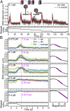
Overwinding of DNA by reverse gyrase leads to indefinite rotations. (A) Sequential images at 132-ms intervals of a fluorescent daughter bead attached to a tethered magnetic bead. White dots show the center of rotation. View from above in Fig. 1D. The image size is 2.8 × 2.8 μm2. The images are from the yellow portion of the red curve in B. Also see Movie S1. (B) Time courses of bead rotation at 10 nM reverse gyrase and monitored sample temperature. Thick straight lines show the linear fit to the portion within ±1 °C of 50 °C or 71 °C, from which the average rotary speed is calculated. (Inset) Temperature dependence of the average rotary speeds at 10 nM reverse gyrase under DNA tension of 0.5 pN. Numbers are n (beads examined; approximately twice the number of beads for 50 °C, where the second measurement occasionally failed), and error bars show SD.
Fig. 3.
Fig. 3.
Concentration dependence of overwinding activity at 0.5 pN of tension. (A and B) Rotation time courses at two temperatures. Colored numbers show [reverse gyrase] in nM. (C and D) [Reverse gyrase] dependence of the rotary speed (C) and the probability of rotation (D). Curves in A and B, with additional data to include all beads that satisfied conditions iiii in the main text, are each fitted with a straight line to give individual rotary speeds, which are classified as rotating (>0.01 rps) or nonrotating (<0.01 rps). The average speed of rotating beads is shown in C, with error bars showing SD. Numbers in D show the total beads analyzed (71 °C) or approximately twice the number of beads (50 °C; data from 50-60-50 °C measurements are included).
Fig. 4.
Fig. 4.
Plectoneme formation by reverse gyrase. (A) Repetitive bead sinking in response to unwinding magnet rotation at 0.1 nM reverse gyrase. Unwinding magnet turns are taken as negative. After several trials under the same tension, we rotated the magnets in the overwinding direction to let the bead sink close to the surface (end of the curve). We then changed the tension and repeated the same procedure. (B, Left) Sinking time courses at various tensions for the bead in A. Individual time courses distinguished by colors are first 1-s median-filtered (smoother curves), aligned at the end of unwinding rotation (time 0), and averaged (thick cyan curve). At ≤0.2 pN, where sinking is significant, we fit the average with the magenta line between 0 s and the midpoint of sinking, with the final level (magenta dashed line) being estimated at 24–29 s; at ≥0.3 pN, the fit is between 0 and 10 s. Magenta numbers show estimated overwinding rates. (B, Right) Bead sinking by magnet rotation in the absence of reverse gyrase. Magenta lines show linear fit (also see Fig. S3).
Fig. 5.
Fig. 5.
Overwinding activity plotted against DNA tension F (A) and torque Γ required for plectoneme growth (B). Dots represent individual beads; circles represent their average, with error bars showing SD; and squares represent free rotation results from Fig. 3C. The dashed line in A shows the fit with exp(−Fδ/kBT).
Fig. 6.
Fig. 6.
DNA-dependent ATPase activity of reverse gyrase at 71 °C. Reverse gyrase (10 nM) was incubated with 5 mM ATP and 100 nM DNA (147-nt ssDNA or dsDNA containing a bubble). Each time course with a different symbol was fitted with a line to obtain an individual rate, although averaged lines are shown here. The hydrolysis rate without enzyme was 2.3 ± 0.1 μM⋅min−1 (n = 3). This value was subtracted from other rates to give the ATPase activity of reverse gyrase of 20 ± 3 s−1 with bubbled DNA (n = 3), 4.8 s−1 with ssDNA (n = 1), and 0.0 ± 0.3 s−1 without DNA (n = 2).
Fig. 7.
Fig. 7.
Chiral operation by reverse gyrase. (AE) Presumed mechanism of passive strand passage in topo IA. The magenta strand is held (hatched) in a groove. (A) Catalytic tyrosine (red circle) cuts the strand and binds one end. (B) Thermal opening of the gate to the central cavity allows thermal strand passage in either direction (light or dark green). (C) When the gate closes and the cut strand happens to be religated, the Lk has changed by one (ΔLk = ±1). (D) Reopening of the gate without strand scission allows the green strand to exit from the cavity. The ΔLk localized in the ss region is spread (diluted) into the entire DNA through rotation of the ss/ds junction. (E) Enzyme is then ready for another passage. All events are stochastic and reversible. (FJ) In reverse gyrase, gate opening is likely controlled by ATP hydrolysis in the helicase domain (light blue) through the latch domain (light green). Biased strand passage may occur as follows. The ds/ss junction on the scissile side (F, Left) and a distant part of the green strand (F, Right) are both bound to the enzyme. (G) Coupled to unlocking of gate motion, the ds region elongates by base pairing, rotating the two ss ends to bias the passage correctly. Religation occurs while the bias is effective (H), and the green strand exits with ΔLk = +1 (I). The gate is locked, and DNA is allowed to rotate to export the ΔLk (I→J). The export would fail if the DNA were already highly overwound. Many variations of this scheme are possible.

References

    1. Kikuchi A, Asai K. Reverse gyrase—A topoisomerase which introduces positive superhelical turns into DNA. Nature. 1984;309(5970):677–681. - PubMed
    1. Suzuki T, et al. Sulfolobus tokodaii sp. nov. (f. Sulfolobus sp. strain 7), a new member of the genus Sulfolobus isolated from Beppu Hot Springs, Japan. Extremophiles. 2002;6(1):39–44. - PubMed
    1. Forterre P, Mirambeau G, Jaxel C, Nadal M, Duguet M. High positive supercoiling in vitro catalyzed by an ATP and polyethylene glycol-stimulated topoisomerase from Sulfolobus acidocaldarius. EMBO J. 1985;4(8):2123–2128. - PMC - PubMed
    1. Nakasu S, Kikuchi A. Reverse gyrase; ATP-dependent type I topoisomerase from Sulfolobus. EMBO J. 1985;4(10):2705–2710. - PMC - PubMed
    1. Serre MC, Duguet M. Enzymes that cleave and religate DNA at high temperature: The same story with different actors. Prog Nucleic Acid Res Mol Biol. 2003;74:37–81. - PubMed

Publication types

LinkOut - more resources