. 2015 Jan;28(1):11-20.
doi: 10.1293/tox.2014-0036. Epub 2014 Sep 22.

Mead acid supplementation does not rescue rats from cataract and retinal degeneration induced by N-methyl-N-nitrosourea

Affiliations

Mead acid supplementation does not rescue rats from cataract and retinal degeneration induced by N-methyl-N-nitrosourea

Yuko Emoto et al. J Toxicol Pathol. 2015 Jan.

Erratum in

  • Errata (Printer's correction).
    [No authors listed] [No authors listed] J Toxicol Pathol. 2016 Jan;29(1):74. Epub 2016 Feb 17. J Toxicol Pathol. 2016. PMID: 26989306 Free PMC article.

Abstract

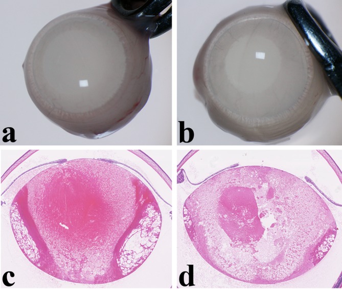
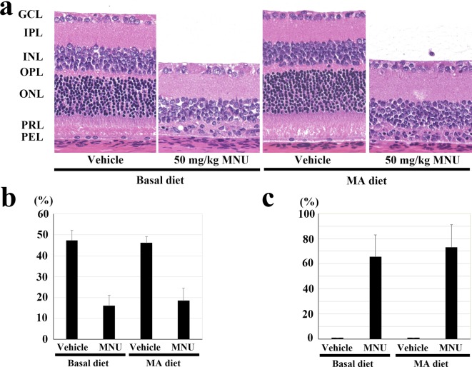
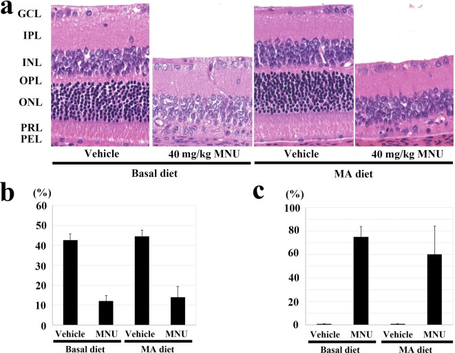
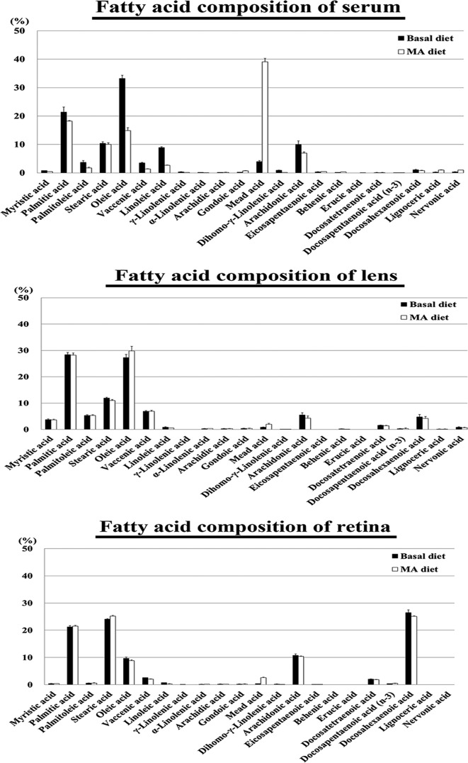
Fatty acids and their derivatives play a role in the response to ocular disease. Our current study investigated the effects of dietary mead acid (MA, 5,8,11-eicosatrienoic acid) supplementation on N-methyl-N-nitrosourea (MNU)-induced cataract and retinal degeneration in Sprague-Dawley rats. Experiment 1 was designed to inhibit cataract formation, with the dams fed a 2.4% MA or basal (<0.01% MA) diet during lactational periods. On postnatal day 7, male pups received a single intraperitoneal (ip) injection of 50 mg/kg MNU or vehicle. Lens opacity and morphology were examined 7 and 14 days after the MNU injection. Experiment 2 was designed to inhibit retinal degeneration and was performed with female postweaning rats. In this experiment, dams were fed the 2.4% MA or basal diet during the lactational periods. Thereafter, the female pups were continuously fed the same diets during their postweaning periods. On postnatal day 21 (at weaning), pups received a single ip injection of 50 mg/kg MNU. Retinal morphology was examined 7 days after the MNU injection. In experiment 3, six-week-old female rats were fed the 2.4% MA or basal diet starting at one week before the MNU injection and were then continuously fed the same diets until sacrifice. Rats at 7 weeks of age were given a single ip injection of 40 mg/kg MNU, and the retina was then examined morphologically one week after the MNU injection. In experiment 1, mature cataract was found in all of the MNU-treated groups, with or without MA supplementation. In experiments 2 and 3, atrophy of both the peripheral and central outer retina occurred in all rats exposed to MNU, with or without MA supplementation, respectively. The severities of the cataracts and retinal atrophy in the rats were similar regardless of MA supplementation. Dietary mead acid, which is used as a substitute in essential fatty acid deficiency in the body, does not modify MNU-induced cataract and retinal degeneration in rat models.

Keywords: N-methyl-N-nitrosourea; arachidonic acid; cataract; mead acid; rats; retinal degeneration.


Conflict of interest statement

Declaration of Conflicting Interests: The authors declare that they have no competing interests.

Figures

Fig. 1.
Fig. 1.
Schema of the experimental protocol. Experiment 1 was designed to inhibit cataract formation in the MNU-induced cataract model using male neonatal rats. Experiments 2 and 3 were designed to confirm the effects on the MNU-induced retinal degeneration models using female postweaning and adult rats, .
Fig. 2.
Fig. 2.
Lens opacity and mature cataract in experiment 1. (a) A severe degree of opacity in the whole lens is seen two weeks after injection of N-methyl-N-nitrosourea (MNU) in a rat fed a basal diet. (b) Similar lens opacity is seen in an MNU-treated rat fed a 2.4% mead acid (MA) diet. (c) Histopathologically, swelling, vacuolation and liquefied fibers occur in the whole lens at two weeks after injection of MNU in a rat fed a basal diet. These changes corresponded to the diagnosis of mature cataract. (d) Mature cataract is similarly seen in the whole lens of a MNU-treated rat fed an MA diet. Hematoxylin and eosin (HE) staining, ×30.
Fig. 3.
Fig. 3.
Effect of the mead acid (MA) diet on 50 mg/kg N-methyl-N-nitrosourea (MNU)-induced retinal damage in the postweaning rats in experiment 2. (a) Histology of the central retina in rats with or without MNU that were fed 2.4% MA and basal diets. At 7 days after a single ip injection of MNU, the outer nuclear layer and photoreceptor layer disappeared in the rats fed the MA and basal diets. GCL, ganglion cell layer; IPL, inner plexiform layer; INL, inner nuclear layer; OPL, outer plexiform layer; ONL, outer nuclear layer; PRL, photoreceptor cell layer; and PEL, pigment epithelial cell layer. HE staining, ×200. (b) Photoreceptor cell ratio in the central retina 7 days after a single ip injection of MNU in rats fed 2.4% MA and basal diets. Rats treated with 50 mg/kg MNU and fed MA the diet do not have any statistically significant changes in their photoreceptor ratios at the central retina compared with the MNU-treated basal diet group. The index is calculated as [(outer retinal thickness / total retinal thickness) ×100]. (c) Retinal damage ratio in MNU-treated rats fed 2.4% MA and basal diets. Rats fed the MA diet do not show any statistically significant changes in the retinal damage ratio compared with the MNU-treated basal diet group. The index was evaluated as [(length of retina composed of less than four photoreceptor cells / whole retinal length) × 100]. The mean ± SE of the five rats in each treatment group is shown.
Fig. 4.
Fig. 4.
Effect of the mead acid (MA) diet on 40 mg/kg N-methyl-N-nitrosourea (MNU)-induced retinal damage in adult rats in experiment 3. (a) Histology of the central retina in rats with or without MNU who were fed the 2.4% MA and basal diets. At 7 days after a single ip injection of MNU, the outer nuclear layer and photoreceptor layer disappeared in the rats fed the MA and basal diets. GCL, ganglion cell layer; IPL, inner plexiform layer; INL, inner nuclear layer; OPL, outer plexiform layer; ONL, outer nuclear layer; PRL, photoreceptor cell layer; and PEL, pigment epithelial cell layer. HE staining, ×200. (b) Photoreceptor cell ratio in the central retina 7 days after a single ip injection of MNU in rats fed the 2.4% MA and basal diets. MNU-treated rats fed the MA diet have a statistically significant decrease in their photoreceptor ratio at the central retina, which is similar to that seen in the MNU-treated basal diet group. The index is calculated as [(outer retinal thickness / total retinal thickness) ×100]. (c) Retinal damage ratio in MNU-treated rats fed the 2.4% MA and basal diets. Rats fed the MA diet do not show any statistically significant changes in the retinal damage ratio, which is similar to that seen in the MNU-treated basal diet group. The index is evaluated as [(length of retina composed of less than four photoreceptor cells / whole retinal length) ×100]. The mean ± SE of the six or seven rats in each treatment group is shown.
Fig. 5.
Fig. 5.
Fatty acid composition of the serum, lens and retina of the vehicle-treated rats. In the serum of the rats fed the 2.4% mead acid (MA) diet, the MA composition of the total fatty acids was higher than that seen in the serum of the rats fed the basal diet. In the lens and retina of the rats fed the 2.4% (MA) diet, although the MA composition was higher, the differences were not remarkable when compared with the serum values. The mean ± SE of four rats in the basal group and three rats in the MA diet group are shown.

References

    1. World Health Organization (WHO), Prevention of Blindness and Deafness Programme Global Data on Visual Impairments 2010. 2012. Retrieved July 15, 2014, from World Health Organization Web site: http://www.who.int/blindness/GLOBALDATAFINALforweb.pdf.
    1. Resnikoff S, and Pararajasegaram R. Blindness prevention programmes: past, present, and future. Bull World Health Organ. 79: 222–226 2001. - PMC - PubMed
    1. Hartong DT, Berson EL, and Dryja TP. Retinitis pigmentosa. Lancet. 368: 1795–1809 2006. - PubMed
    1. Berson EL, Rosner B, Sandberg MA, Weigel-DiFranco C, and Willett WC. ω-3 intake and visual acuity in patients with retinitis pigmentosa receiving vitamin A. Arch Ophthalmol. 130: 707–711 2012. - PMC - PubMed
    1. Tsubura A, Yuri T, Yoshizawa K, Uehara N, and Takada H. Role of fatty acids in malignancy and visual impairment: epidemiological evidence and experimental studies. Histol Histopathol. 24: 223–234 2009. - PubMed

LinkOut - more resources