. 2015 May 29;10(5):e0127794.
doi: 10.1371/journal.pone.0127794. eCollection 2015.

Studying Cat (Felis catus) Diabetes: Beware of the Acromegalic Imposter

Affiliations

Studying Cat (Felis catus) Diabetes: Beware of the Acromegalic Imposter

Stijn J M Niessen et al. PLoS One. .

Abstract

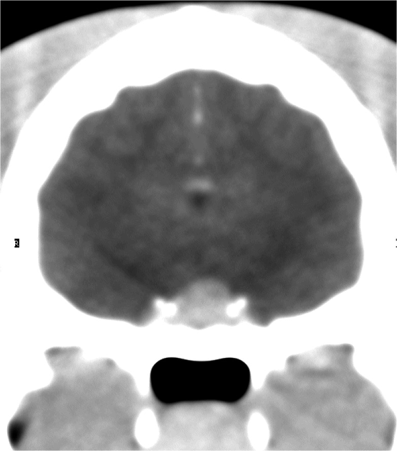
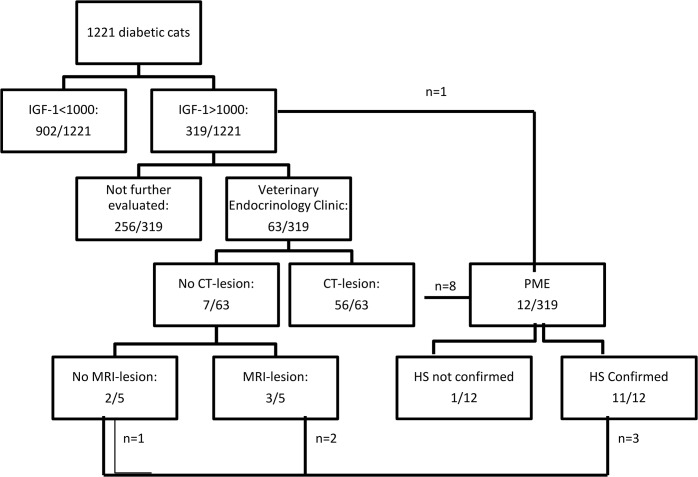
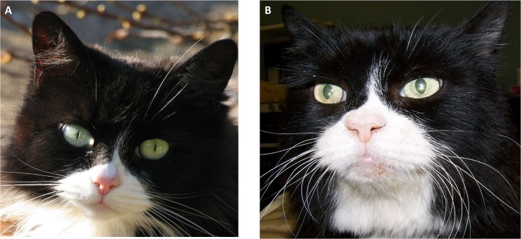
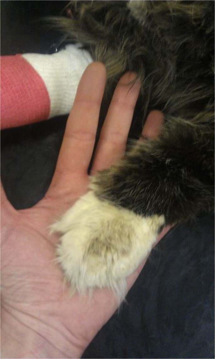
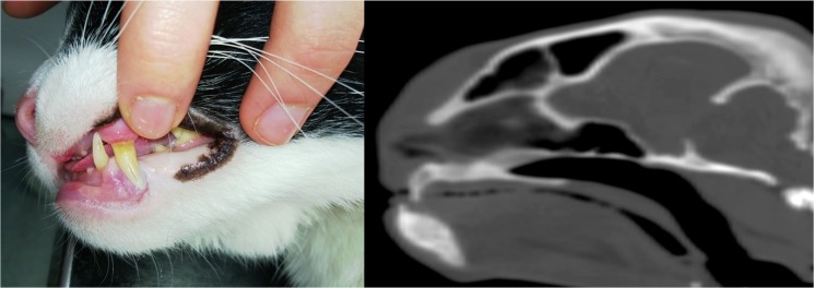
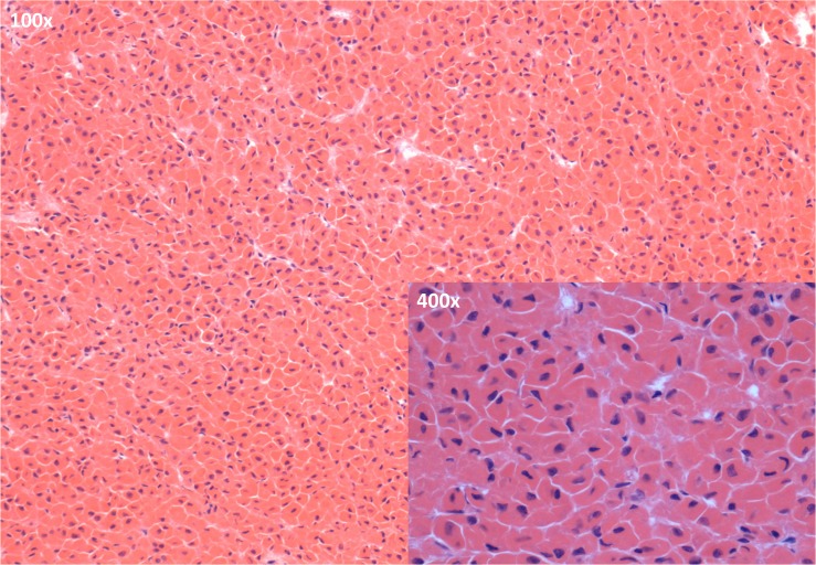
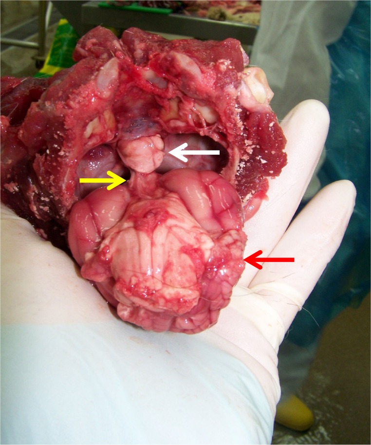
Naturally occurring diabetes mellitus (DM) is common in domestic cats (Felis catus). It has been proposed as a model for human Type 2 DM given many shared features. Small case studies demonstrate feline DM also occurs as a result of insulin resistance due to a somatotrophinoma. The current study estimates the prevalence of hypersomatotropism or acromegaly in the largest cohort of diabetic cats to date, evaluates clinical presentation and ease of recognition. Diabetic cats were screened for hypersomatotropism using serum total insulin-like growth factor-1 (IGF-1; radioimmunoassay), followed by further evaluation of a subset of cases with suggestive IGF-1 (>1000 ng/ml) through pituitary imaging and/ or histopathology. Clinicians indicated pre-test suspicion for hypersomatotropism. In total 1221 diabetic cats were screened; 319 (26.1%) demonstrated a serum IGF-1>1000 ng/ml (95% confidence interval: 23.6-28.6%). Of these cats a subset of 63 (20%) underwent pituitary imaging and 56/63 (89%) had a pituitary tumour on computed tomography; an additional three on magnetic resonance imaging and one on necropsy. These data suggest a positive predictive value of serum IGF-1 for hypersomatotropism of 95% (95% confidence interval: 90-100%), thus suggesting the overall hypersomatotropism prevalence among UK diabetic cats to be 24.8% (95% confidence interval: 21.2-28.6%). Only 24% of clinicians indicated a strong pre-test suspicion; most hypersomatotropism cats did not display typical phenotypical acromegaly signs. The current data suggest hypersomatotropism screening should be considered when studying diabetic cats and opportunities exist for comparative acromegaly research, especially in light of the many detected communalities with the human disease.


Conflict of interest statement

Competing Interests: The authors have declared that no competing interests exist.

Figures

Fig 1
Fig 1. Protrusion of the pituitary beyond the dorsal rim of the sella turcica.
This figure shows an example of a transverse, post-contrast CT image in a cat with HS. In this instance the dorsoventral dimension (height) of the enlarged pituitary was 6mm.
Fig 2
Fig 2. Overview of the screening results.
In total 1221 cats with diabetes mellitus were screened for HS; the results are summarised in this figure. IGF-1: insulin-like growth factor-1; CT: computed tomography; MRI: magnetic resonance imaging; HS: hypersomatotropism; n: number of animals; PME: post-mortem evaluation.
Fig 3
Fig 3. Example of a cat before and after onset of HS-induced changes.
Photo a was taken in 2009 and photo b after onset of HS-induced changes in 2012. HS-induced broad facial features are evident in the later picture.
Fig 4
Fig 4. Paws of an overtly acromegalic cat.
This figure shows a cat with confirmed acromegaly demonstrating so-called clubbed paws; enlargement of the paws due to acromegaly.
Fig 5
Fig 5. Example of prognathia inferior.
This figure shows a cat with confirmed acromegaly demonstrating prognathia inferior (a), including a sagittal CT view of the cat’s head illustrating this conformational change further (b).
Fig 6
Fig 6. Radiograph of the chest and abdomen of an overtly acromegalic cat.
Cranial abdominal organomegaly was demonstrated by this radiographic view of the chest and abdomen of a diabetic cat subsequently diagnosed with HS.
Fig 7
Fig 7. Microscopic image of the anterior pituitary of a diabetic cat with confirmed HS.
A clear predominance of acidophils is apparent in the sections of the pars distalis of the anterior pituitary (H&E staining; main image: objective 100x; inset: objective: 400x)
Fig 8
Fig 8. Macroscopic necropsy picture of the brain of a diabetic cat with confirmed HS.
This picture shows the ventral aspect of the brain of a cat with HS as it was removed from the cranial cavity and a clearly enlarged pituitary falling out of the sella turcica (cranium is held upside down; white arrow: pituitary gland; yellow arrow: pituitary stalk; red arrow: cerebrum). Histopathology confirmed the presence of an acidophilic macroadenoma.

References

    1. McCann TM, Simpson KE, Shaw DJ, Butt JA, Gunn-Moore DA. Feline diabetes mellitus in the UK: the prevalence within an insured cat population and a questionnaire-based putative risk factor analysis. J Feline Med Surg. 2007; 9(4): 289–299. - PMC - PubMed
    1. Lederer R, Rand JS, Jonsson NN, Hughes IO, Morton JM. Frequency of feline diabetes mellitus and breed predisposition in domestic cats in Asutralia. Vet J. 2009;179(2): 254–258. - PubMed
    1. Prahl AL, Guptill L, Glickman NW, Tetrick M, Glickman LTJ. Time trends and risk factors for diabetes mellitus in cats presented to veterinary teaching hospitals. J Feline Med Surg. 2007; 9(5): 351–358. - PMC - PubMed
    1. Dirtu AC, Niessen SJ, Jorens PG, Covaci A. Organohalogenated contaminants in domestic cats’ plasma in relation to spontaneous acromegaly and type 2 diabetes mellitus: A clue for endocrine disruption in humans? Environ Int. 2013; 57–58: 60–67. 10.1016/j.envint.2013.10.018 - DOI - PubMed
    1. Hoenig M. Comparative aspects of diabetes mellitus in dogs and cats. Mol Cell Endocrinol. 2002; 197(1–2): 221–229. - PubMed

Substances