. 2015 May 29;11(5):e1004932.
doi: 10.1371/journal.ppat.1004932. eCollection 2015 May.

Vaccine-Elicited Tier 2 HIV-1 Neutralizing Antibodies Bind to Quaternary Epitopes Involving Glycan-Deficient Patches Proximal to the CD4 Binding Site

Affiliations

Vaccine-Elicited Tier 2 HIV-1 Neutralizing Antibodies Bind to Quaternary Epitopes Involving Glycan-Deficient Patches Proximal to the CD4 Binding Site

Ema T Crooks et al. PLoS Pathog. .

Abstract

Eliciting broad tier 2 neutralizing antibodies (nAbs) is a major goal of HIV-1 vaccine research. Here we investigated the ability of native, membrane-expressed JR-FL Env trimers to elicit nAbs. Unusually potent nAb titers developed in 2 of 8 rabbits immunized with virus-like particles (VLPs) expressing trimers (trimer VLP sera) and in 1 of 20 rabbits immunized with DNA expressing native Env trimer, followed by a protein boost (DNA trimer sera). All 3 sera neutralized via quaternary epitopes and exploited natural gaps in the glycan defenses of the second conserved region of JR-FL gp120. Specifically, trimer VLP sera took advantage of the unusual absence of a glycan at residue 197 (present in 98.7% of Envs). Intriguingly, removing the N197 glycan (with no loss of tier 2 phenotype) rendered 50% or 16.7% (n = 18) of clade B tier 2 isolates sensitive to the two trimer VLP sera, showing broad neutralization via the surface masked by the N197 glycan. Neutralizing sera targeted epitopes that overlap with the CD4 binding site, consistent with the role of the N197 glycan in a putative "glycan fence" that limits access to this region. A bioinformatics analysis suggested shared features of one of the trimer VLP sera and monoclonal antibody PG9, consistent with its trimer-dependency. The neutralizing DNA trimer serum took advantage of the absence of a glycan at residue 230, also proximal to the CD4 binding site and suggesting an epitope similar to that of monoclonal antibody 8ANC195, albeit lacking tier 2 breadth. Taken together, our data show for the first time that strain-specific holes in the glycan fence can allow the development of tier 2 neutralizing antibodies to native spikes. Moreover, cross-neutralization can occur in the absence of protecting glycan. Overall, our observations provide new insights that may inform the future development of a neutralizing antibody vaccine.


Conflict of interest statement

The authors have declared that no competing interests exist. Some authors are employed with Altravax Inc, but this does not affect competing interests. No employment, consultancies, patents or products in development or on market represent a competing interest with this article. This does not alter our adherence to all PLOS Pathogens policies on sharing data and materials.

Figures

Fig 1
Fig 1. Overview of human plasmas and animal sera.
Our plasma and serum panel consisted of human plasmas from 4 HIV-1-infected donors and one uninfected donor, a pool of rabbit sera generated against monomeric JR-FL gp120, groups of rabbit and guinea pig sera generated to various JR-FL Env-VLP-based immunogens. Group 5 animals all received JR-FL-based immunogens via different DNA prime-protein boost protocols. The absolute dose of gp120 and gp140 immunogens is shown. VLP doses are given in estimated gp120 equivalents [28]. Adjuvants and immunization routes are shown (abbreviations are: IM intramuscular, SC subcutaneous and ID intradermal).
Fig 2
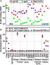
Fig 2. Serum binding and neutralization profiles.
A) ELISA titers using recombinant JR-FL gp120, gp41 and bald-VLPs. Data shown are means of at least two repeats. B) Neutralization activity was measured in two different assay formats. TZM-bl cells were used to measure nAbs against the JR-FL WT E168K and SIVmac239 viruses in a "leave in" format. CF2.ThCD4.CCR5 cells were used to measure activity against the JR-FL SOS E168K virus in a “washout” format. All neutralization assays were performed at least three times in duplicate. Mean titers and standard deviations are shown.
Fig 3
Fig 3. Serum recognition of the native trimer and effects of a key mutation in the CD4 binding loop.
MAbs and serum binding to the native Env trimer was assessed in BN-PAGE shift assays. Briefly, mAbs (30μg/ml) or sera (1:2 dilution) were incubated with protease-digested 2nd generation A) JR-FL SOS E168K VLPs or B) JR-FL SOS E168K+D368R VLPs. The VLPs were then washed and lysed, and Env was resolved by BN-PAGE-Western blot. Ferritin was used as a molecular weight marker. The unliganded trimer is indicated by a cartoon. The abbreviation “UND VLP” (immunogen for rabbit 617) refers to “undigested VLPs”.
Fig 4
Fig 4. Adsorption to native Env trimer expressed on cell surfaces removes potent serum neutralizing activities.
IgG was first protein A-purified from sera and adjusted to the original serum concentration (~10mg/ml). Samples were then adsorbed repeatedly to native Env trimer-expressing cells. IgG was then re-isolated and adjusted to the original volume. A) Pre- and post-adsorption IgG samples were then assessed for neutralizing activity against JR-FL gp160∆CT WT E168K in the TZM-bl assay. B) Purified IgG from serum 613 was monitored before and after adsorption by silver stain. Each sample was titrated.
Fig 5
Fig 5. Potent vaccine sera target epitopes that overlap the CD4 binding site.
Selected vaccine sera (1:10 dilution) were competed against various biotinylated monoclonal bnAbs for binding to SOS E168K trimer VLPs by ELISA. E168K+N189A mutant trimer VLPs were used for assays involving biotinylated V2 quaternary mAbs to ensure a full epitope knock in [56]. Three previously mapped HIV-1-infected plasmas were included for reference purposes [54]. These are (epitopes in parentheses) 1702 (some MPER and unmapped nAb), 1686 (predominantly CD4bs nAb) and BB34 (predominantly MPER nAb). Numbers are expressed as the residual % binding titer of biotinylated mAb in the presence of competitor compared to its titer in the presence of a control rabbit prebleed or HIV-1 seronegative human plasma (from donor 210). Each assay was performed at least in duplicate. Standard errors are shown in S8 Fig.
Fig 6
Fig 6. Domain and residue-level mapping of potent rabbit sera.
A) Domains that determine the neutralizing activities of each of the 4 sera shown were inferred from the chimera analysis in S11 Fig. Domains are color-coded: purple indicates JR-FL Env domains, salmon indicates JR-CSF Env domains. B) C2 domains of JR-FL and JR-CSF Envs were aligned. To save space, some conserved residues are abbreviated as dots. Eight differences in the JR-CSF sequence are shown in yellow. The conservation of JR-CSF residues at each of these positions is shown. A disulfide-bonded hairpin is shown by red lines. The sensitivities of the 613 and 647 sera to JR-CSF mutations are shown in green, C) 613 and 647 serum neutralization ID50s of JR-FL clones bearing JR-CSF-based residue substitutions. Each mutant was also evaluated for neutralization sensitivity (IC50s in μg/ml) to mAbs b12 and CO11.
Fig 7
Fig 7. Serum 613 neutralization breadth of N197 knockout mutants.
613 serum ID50s against N197 glycan knockout mutants of various clade B (blue symbols) and non-clade B (green symbols) in the TZM-bl assay. N197 glycan knockout mutants sensitive to 613 serum are indicated. Raw data are shown in S5 Table.
Fig 8
Fig 8. Fingerprint analysis of the 613 serum suggests weak associations with CD4 binding site and V2 quaternary epitopes.
A fingerprint analysis [69] was performed based on the sequences of neutralized and non-neutralized N197 mutants of a subset of 24 mutants and 12 bnAb specificities (see methods). The selected N197 mutants (the parent Env Genbank accession number in parentheses) were 6101.10 (AAT36747), 7165 (AAW64252), 92US715 (AAB04079), AC10.29 (AAW64261), ADA.DG (AAR05843), BG1168 (AAW64258), BL01.DG (AAN39728), CNE12 (ADI62512), CNE4 (ADI62567), JR-CSF (AAB03749), JR-FL (AAB05604), PVO.04 (AAW64259), Q769.d22 (AAM66234), Q769.h5 (AAM66238), REJO4541.67 (AAW64264), RHPA.7 (AAW64262), TRJO (AAW64265), TRO.11 (AAW64260), WITO.33 (AAW64266), YU2.DG (M93258), ZA012.29 (ACF75939), ZM106.9 (AAR09562), ZM55.28a (AAR09381) and QH0515 (AAW64255). Delineation values >0.2 are considered to indicate the presence of particular specificities in the polyclonal serum.
Fig 9
Fig 9. Modeling the epitope footprints of neutralizing sera 613 and 647.
The binding sites of the two potently neutralizing rabbit sera 613 and 647 are modeled on BG505 SOSIP.664 Env trimers. Images were created from pdb 4TVP and show side and top views, as indicated. Gp41 is shown in metallic grey. Different gp120 protomers are shown in different shades of green so they can be discerned from one another. In nature, a significant proportion of glycans are complex in nature and may exhibit branches that are difficult to predict and/or unequivocally model [89]. Therefore, all glycans (mostly shown in dark blue) were rendered as Man8GlcNAc2. Since complex glycans usually have a greater mass than Man8GlcNAc2 (see Fig 1 of [64]), the glycan coverage shown here may be a slight underestimate compared to infectious spikes. On the other hand, this model employs all available sequons on the trimer, whereas in nature, close proximity may result in some sequons being unused [64]. A) The CD4 binding loop (residues 360–370) and other putative CD4 contacts at positions 427 and 477 are shaded yellow. A putative "glycan fence", consisting of glycans N197, N276, N362 and N386 is shown in red (for N197) and magenta. The N160 glycan and K169 residue contacts of PG cluster (V1V2 loop-specific) mAbs at the apex of the trimer are shown in orange. B) Fab VRC01 (3NGB), rendered in pea green ribbon format, is shown docked to the trimer. C) The N197 glycan is removed and the underlying N197 residue is labeled red. D) The putative binding site for the 613 serum rendered as a cyan Fab is modeled. E) Residues 230, 238 (loop A) and 269 (loop C) of the gp120 C2 region targeted by serum 647 are shown in red. The N234 glycan is omitted, as in the JR-FL strain. However, residue N241 is colored in black to indicate an additional glycan in the JR-FL strain that is absent in BG505. F) Fab 8ANC195 is docked on the structure in part E, as the best approximation of the 647 serum. G) The N234 glycan present in BG505 is added back, showing how it could clash with 647 nAbs.

References

    1. Schiffner T, Sattentau QJ, Dorrell L (2013) Development of prophylactic vaccines against HIV-1. Retrovirology 10: 72 10.1186/1742-4690-10-72 - DOI - PMC - PubMed
    1. Mascola JR, Montefiori DC (2010) The role of antibodies in HIV vaccines. Annu Rev Immunol 28: 413–444. 10.1146/annurev-immunol-030409-101256 - DOI - PubMed
    1. Crooks ET, Jiang P, Franti M, Wong S, Zwick MB, et al. (2008) Relationship of HIV-1 and SIV envelope glycoprotein trimer occupation and neutralization. Virology 377: 364–378. 10.1016/j.virol.2008.04.045 - DOI - PMC - PubMed
    1. Kwong PD, Mascola JR (2012) Human Antibodies that Neutralize HIV-1: Identification, Structures, and B Cell Ontogenies. Immunity 37: 412–425. 10.1016/j.immuni.2012.08.012 - DOI - PMC - PubMed
    1. Pancera M, Zhou T, Druz A, Georgiev IS, Soto C, et al. (2014) Structure and immune recognition of trimeric pre-fusion HIV-1 Env. Nature 514: 455–461. 10.1038/nature13808 - DOI - PMC - PubMed

Publication types

MeSH terms