. 2015 Aug 7;6(22):18800-18.
doi: 10.18632/oncotarget.4033.

Differential modulation of AMPK/PPARα/UCP2 axis in relation to hypertension and aging in the brain, kidneys and heart of two closely related spontaneously hypertensive rat strains

Affiliations

Differential modulation of AMPK/PPARα/UCP2 axis in relation to hypertension and aging in the brain, kidneys and heart of two closely related spontaneously hypertensive rat strains

Speranza Rubattu et al. Oncotarget. .

Abstract

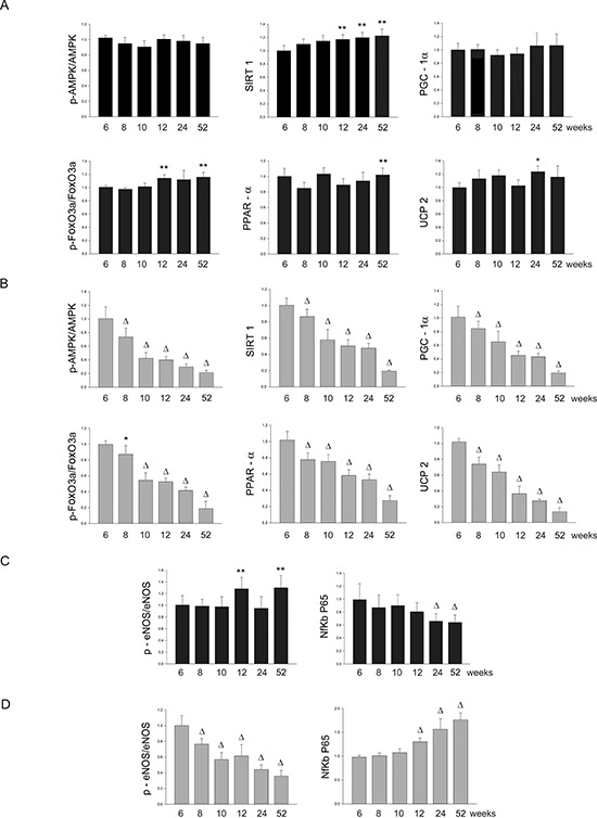
Objectives: We examined expression protein of AMPK/SIRT1/PGC1α/PhoxO3a/PPARα/UCP2 pathway in brain, kidneys and heart of stroke-prone spontaneously hypertensive rat (SHRSP) vs stroke-resistant SHR (SHRSR) at different weeks of age, up to one year, in order to test the hypothesis that abnormalities within this pathway could associate with higher susceptibility of SHRSP to develop hypertension-related vascular damage.

Background: SHRSP develops severe hypertension and related target organ damage. Marked reduction of uncoupling protein 2 (UCP2) expression upon high salt-low potassium diet associates with increased renal injury in SHRSP. UCP2 may represent a key mitochondrial protein involved in cardiovascular damage.

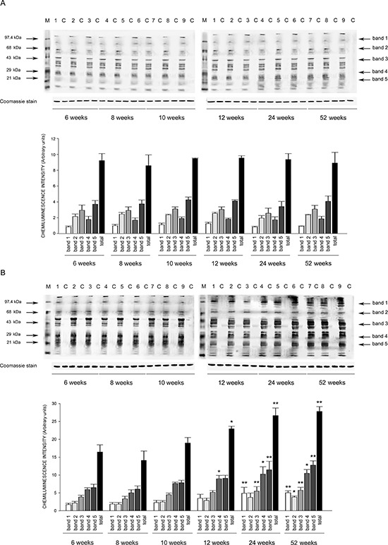
Results: At 2 months of age a significant down-regulation of UCP2 expression at both mRNA and protein levels was found, along with reduced protein expression of all components of UCP2 regulatory pathway, in tissues of SHRSP but not of SHRSR, that progressed with hypertension development and aging. A significant increase of both oxidative stress and inflammation was detected in tissues of SHRSP as a function of age. SBP levels were significantly higher in SHRSP than SHRSR at 3 months of age and thereafter. At one year of age, higher degree of renal damage, with proteinuria and severe glomerular and tubulo-interstitial fibrosis, of cerebral damage, with significant vessel extravasation and stroke occurrence, and of myocardial damage was detected in SHRSP than SHRSR.

Conclusions: The early significant reduced expression of the antioxidant AMPK/PPARα/UCP2 pathway that progressed throughout lifetime may contribute to explain higher predisposition of SHRSP to oxidative stress dependent target organ damage in the context of severe hypertension.

Keywords: Gerotarget; SHRSP; aging; hypertension; target organ damage; uncoupling protein 2.


Conflict of interest statement

CONFLICTS OF INTEREST

None to be declared.

Figures

Figure 1
Figure 1. Panel A: western blots of AMPK/SIRT1/PGC1α/PhoxO3a/PPARα/UCP2 pathway throughout lifetime in the brains of SHRSR and SHRSP
Panel B: western blots of phosphoeNOS/eNOS and of NfKb in the brains of the two strains.
Figure 2
Figure 2. Panel A: densitometric analysis of each component of the AMPK/SIRT1/PGC1α/PhoxO3a/PPARα/UCP2 pathway at different weeks of age in the brains of SHRSR
Panel B: same analysis in SHRSP; Panel C: densitometric analysis of phosphoeNOS/eNOS and of NfKb in SHRSR; Panel D: same analysis in SHRSP. **p < 0.01 vs 6 weeks of age; Δp < 0.0001 vs 6 weeks of age
Figure 3
Figure 3. Panel A: western blots of AMPK/SIRT1/PGC1α/PhoxO3a/PPARα/UCP2 pathway throughout lifetime in the kidneys of SHRSR and SHRSP
Panel B: western blots of phosphoeNOS/eNOS and of NfKb in the kidneys of the two strains.
Figure 4
Figure 4. Panel A: densitometric analysis of each component of the AMPK/SIRT1/PGC1α/PhoxO3a/PPARα/UCP2 pathway at different weeks of age in the kidneys of SHRSR
Panel B: same analysis in SHRSP; Panel C: densitometric analysis of phosphoeNOS/eNOS and of NfKb in SHRSR; Panel D: same analysis in SHRSP. *p < 0.05 vs 6 weeks of age; **p < 0.01 vs 6 weeks of age; Δp < 0.0001 vs 6 weeks of age
Figure 5
Figure 5. Panel A: western blots of AMPK/SIRT1/PGC1α/PhoxO3a/PPARα/UCP2 pathway throughout lifetime in the heart of SHRSR and SHRSP
Panel B: western blots of phosphoeNOS/eNOS and of NfKb in the heart of the two strains.
Figure 6
Figure 6. Panel A: densitometric analysis of each component of the AMPK/SIRT1/PGC1α/PhoxO3a/PPARα/UCP2 pathway at different weeks of age in the heart of SHRSR
Panel B: same analysis in SHRSP; Panel C: densitometric analysis of phosphoeNOS/eNOS and of NfKb in SHRSR; Panel D: same analysis in SHRSP. *p < 0.05 vs 6 weeks of age; **p < 0.01 vs 6 weeks of age; Δp < 0.0001 vs 6 weeks of age
Figure 7
Figure 7. Western blots of intracellular protein extracts immunostained for carbonylated proteins using the Oxyblot Protein Oxidation Detection kit in the brains of SHRSR and SHRSP
Each lane was loaded with 50 μg of total proteins. Lane M, DNP marker. Each sample is run with its own untreated control (C) Normalization for lane protein loading was performed using Coomassie staining. Bar graphs below the gel blots represent chemiluminenscence intensity relative to the gel loading band. Bands 1–5 refer to the most prominent bands on the blots (identified by arrows), whereas total refer to the total chemiluminescence intensity from all bands. Δp < 0.0001 vs 6 weeks of age
Figure 8
Figure 8. Western blots of intracellular protein extracts immunostained for carbonylated proteins using the Oxyblot Protein Oxidation Detection kit in the kidneys of SHRSR and SHRSP
Each lane was loaded with 50 μg of total proteins. Lane M, DNP marker. Each sample is run with its own untreated control (C) Normalization for lane protein loading was performed using Coomassie staining. Bar graphs below the gel blots represent chemiluminenscence intensity relative to the gel loading band. Bands 1–5 refer to the most prominent bands on the blots (identified by arrows), whereas total refer to the total chemiluminescence intensity from all bands. *p < 0.05 vs 6 weeks of age; Δp < 0.0001 vs 6 weeks of age
Figure 9
Figure 9. Western blots of intracellular protein extracts immunostained for carbonylated proteins using the Oxyblot Protein Oxidation Detection kit in the heart of SHRSR and SHRSP
Each lane was loaded with 50 μg of total proteins. Lane M, DNP marker. Each sample is run with its own untreated control (C) Normalization for lane protein loading was performed using Coomassie staining. Bar graphs below the gel blots represent chemiluminenscence intensity relative to the gel loading band. Bands 1–5 refer to the most prominent bands on the blots (identified by arrows), whereas total refer to the total chemiluminescence intensity from all bands. *p < 0.05 vs 6 weeks of age; **p < 0.01 vs 6 weeks of age
Figure 10
Figure 10. Panel A: representative images of IgG immunostaining detected in the frontal cortex of SHRSR and SHRSP
Panel B: bar graphs represent quantification of number of cerebral blood vessels showing IgG extravasation density for mm2 in 20 randomly selected fields spanning the frontal cortex. *p < 0.05, one year-old SHRSP vs 6 week-old SHRSP; #p < 0.05, one year-old SHRSP vs one year-old SHRSR. Panel C: H&E staining showing the presence of both ischemic and hemorrhagic lesions only in the brain of one year-old SHRSP.
Figure 11
Figure 11. Panel A: representative images of renal arterioles of SHRSR and SHRSP at both 6 weeks and one year of age
Panel B: bar graphs represent values of media-to-lumen area ratio. *p < 0.05, one year-old SHRSP vs 6 week-old SHRSP. Panel C: representative images of glomeruli from kidneys of SHRSR and SHRSP at both 6 weeks and one year of age. Presence of peritubular (<) and perivascular (*) fibrosis is indicated in one year-old rats. Panel D: bar graphs represent quantification of the glomerular percentage area positively stained with PAS. *p < 0.001, one year-old SHRSR vs 6 week-old SHRSR and one year-old SHRSP vs 6 week-old SHRSP; #p < 0.001, one year-old SHRSP vs one year-old SHRSR.
Figure 12
Figure 12. Panel A: representative images of renal fibrosis at both cortical and medullar level in SHRSR and SHRSP at both 6 weeks and one year of age
Presence of interstitial (°), perivascular (*) and glomerular (<) fibrosis is indicated in the cortex of SHRSP rats at one year of age. Panel B: bar graphs represent quantification of fibrosis as percentage of blue area. *p < 0.001, one year-old SHRSP vs 6 week-old SHRSP; #p < 0.001, one year-old SHRSP vs one year-old SHRSR.
Figure 13
Figure 13. Panel A: representative images of intramyocardial coronary arterioles of SHRSR and SHRSP at 6 weeks and one year of age
Panel B: bar graphs represent values of media-to-lumen area ratio. *p < 0.05, one year-old SHRSP vs 6 week-old SHRSP. Panel C: representative images of perivascular and myocardial interstitial collagen accumulation. Panel D: bar graphs represent perivascular collagen area-to-vessel area ratio (PVCA/LA). Panel E: bar graphs represent myocardial interstitial collagen percentage area. *P < 0.05, one year-old SHRSP vs 6 week-old SHRSP.

References

    1. Mattiasson G, Sullivan PG. The emerging functions of UCP2 in heath, disease, and therapeutics. Antioxid Redox Signal. 2006;8:1–38. - PubMed
    1. Moukdar F, Robidoux J, Lyght O, Pi J, Daniel KW, Collins S. Reduced antioxidant capacity and diet-induced atherosclerosis in uncoupling protein-2-deficient mice. J Lipid Res. 2009;50:59–70. - PubMed
    1. Tian XY, Wong WT, Xu A, Lu Y, Zhang Y, Wang L, Cheang WS, Wang Y, Yao X, Huang Y. Uncoupling protein-2 protects endothelial function in diet-induced obese mice. Circ Res. 2012;110:1211–1216. - PubMed
    1. Liu L, Liu J, Tian XY, Wong WT, Lau CW, Xu A, Xu G, Ng CF, Yao X, Gao Y, Huang Y. Uncoupling protein-2 mediates DPP-4 inhibitor-induced restoration of endothelial function in hypertension through reducing oxidative stress. Antioxid Redox Signal. 2014;21:1571–81. - PMC - PubMed
    1. Brand MD. Uncoupling to survive? The role of mitochondrial inefficiency in ageing. Exp Geront. 2000;35:811–820. - PubMed

Publication types

MeSH terms