Review
. 2015 May 27;5(2):1035-62.
doi: 10.3390/biom5021035.

Initial events in bacterial transcription initiation

Affiliations
Review

Initial events in bacterial transcription initiation

Emily F Ruff et al. Biomolecules. .

Abstract

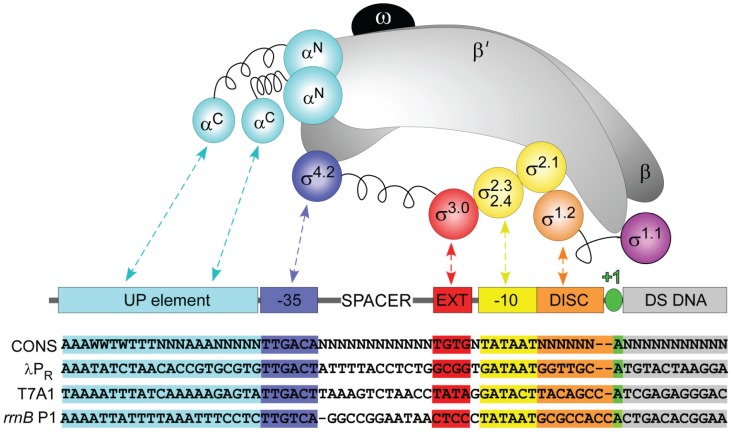
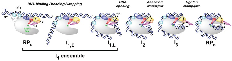
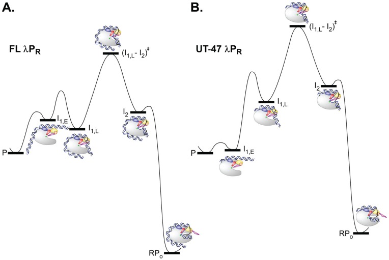
Transcription initiation is a highly regulated step of gene expression. Here, we discuss the series of large conformational changes set in motion by initial specific binding of bacterial RNA polymerase (RNAP) to promoter DNA and their relevance for regulation. Bending and wrapping of the upstream duplex facilitates bending of the downstream duplex into the active site cleft, nucleating opening of 13 bp in the cleft. The rate-determining opening step, driven by binding free energy, forms an unstable open complex, probably with the template strand in the active site. At some promoters, this initial open complex is greatly stabilized by rearrangements of the discriminator region between the -10 element and +1 base of the nontemplate strand and of mobile in-cleft and downstream elements of RNAP. The rate of open complex formation is regulated by effects on the rapidly-reversible steps preceding DNA opening, while open complex lifetime is regulated by effects on the stabilization of the initial open complex. Intrinsic DNA opening-closing appears less regulated. This noncovalent mechanism and its regulation exhibit many analogies to mechanisms of enzyme catalysis.

Keywords: RNA polymerase; kinetics; mechanism; promoter; transcription regulation.


Figures

Figure 1
Figure 1
Sequence-specific interactions between σ70 RNAP and regions of the promoter. Schematic representations of the subunits of RNAP core, σ70, and promoter DNA. RNAP: α2: cyan; β and β': gray; ω: black. σ regions: as shown. Promoter: UP element: cyan; −35 element: blue; extended −10: red; −10 element: yellow; discriminator: orange; transcription start site: green; DNA downstream of the transcription start site: gray. Linker regions in α and σ subunits are shown as springs. Nontemplate strand sequences of a “consensus” and λPR, T7A1 and rrnB P1 promoters are shown below; missing bases are indicated by dashes.
Figure 2
Figure 2
Structural representation of key functional regions of Eσ70 holoenzyme. Structure created from PDB 4LK1, looking down the cleft in (A) and rotated 90° into the page and 90° counterclockwise in (B). Colors of regions of σ are the same as in Figure 1. Additionally shown: β' jaw (β' 1149–1990), brown; β' SI3 (β' insert 6; β' 943–1130), green; β SI1 (β insert 4; β 225–343), pink; active site Mg2+, red ball; αNTD (α 1–234), cyan.
Figure 3
Figure 3
Schematic representation of proposed intermediates in open complex formation and dissociation at λPR promoter. Closed complexes like RPC, I1,E (early), or I1,L (late) can be significant members of the rapidly equilibrating I1 ensemble. The αCTDs are shown in cyan; other colors are the same as in Figure 1.
Figure 4
Figure 4
Mechanism of transcription initiation: resolvable kinetic steps. Minimal mechanisms of open complex formation and dissociation, showing by color coding the steps that contribute to the observed rate constants (kobs, kd) for promoters like λPR that form a stable open complex RPO. For the common experimental situation of excess RNAP, the rate constant for open complex formation (kobs) is determined by the front half of the mechanism (boxed in purple), including the equilibrium constant K1 for formation of the ensemble of closed (I1) intermediates, the forward rate constant k2 of the isomerization step that includes DNA opening, and the excess RNAP concentration. Likewise, kd is determined by the late steps of the mechanism (boxed in green), including the rate constant k-2 for DNA closing and the equilibrium constant K3 for stabilization of the initial open complex I2 to form longer-lived I3 and/or RPO complexes [65,66].
Figure 5
Figure 5
Reaction progress diagrams for open complex formation by (A) FL and (B) UT−47 λPR, interpreted using Mechanism 2. Free energies of the rapidly-equilibrating closed intermediates I1,E and I1,L, the rate- determining transition state (I1,L-I2), and the initial (I2) and stable (RPO) open complexes are calculated relative to promoter DNA (P) for [R] = 30 nM from experimentally-determined values of K1, k2, k-2 and kd for FL λPR [24,60] and UT-47 λPR [75] at 37 °C (see text for details). Free energies of FL and UT-47 λPR are set at the same value. For purposes of illustration, for both variants, we estimate that kopen = 1 s−1 and that (I1,L-I2) decomposition frequencies are 103 s−1 in each direction, and assume that k-2 for UT-47 λPR is the same as that determined for FL λPR [24].

Similar articles

Cited by

References

    1. Decker K.B., Hinton D.M. Transcription regulation at the core: Similarities among bacterial, archaeal, and eukaryotic RNA polymerases. Annu. Rev. Microbiol. 2013;67:113–139. doi: 10.1146/annurev-micro-092412-155756. - DOI - PubMed
    1. Ebright R.H. RNA polymerase: Structural similarities between bacterial RNA polymerase and eukaryotic RNA polymerase II. J. Mol. Biol. 2000;304:687–698. doi: 10.1006/jmbi.2000.4309. - DOI - PubMed
    1. Saecker R.M., Record M.T., Jr., Dehaseth P.L. Mechanism of bacterial transcription initiation: RNA polymerase—Promoter binding, isomerization to initiation-competent open complexes, and initiation of RNA synthesis. J. Mol. Biol. 2011;412:754–771. doi: 10.1016/j.jmb.2011.01.018. - DOI - PMC - PubMed
    1. Dorman C.J. Co-operative roles for DNA supercoiling and nucleoid-associated proteins in the regulation of bacterial transcription. Biochem. Soc. Trans. 2013;41:542–547. doi: 10.1042/BST20120222. - DOI - PubMed
    1. Feklistov A., Sharon B.D., Darst S.A., Gross C.A. Bacterial sigma factors: A historical, structural, and genomic perspective. Annu. Rev. Microbiol. 2014;68:357–376. doi: 10.1146/annurev-micro-092412-155737. - DOI - PubMed

Publication types

MeSH terms

LinkOut - more resources