. 2015 May;56(5):2783-9.
doi: 10.1167/iovs.14-16355.

Effect of internal limiting membrane abrasion on retinal tissues in macular holes

Affiliations

Effect of internal limiting membrane abrasion on retinal tissues in macular holes

David R P Almeida et al. Invest Ophthalmol Vis Sci. 2015 May.

Abstract

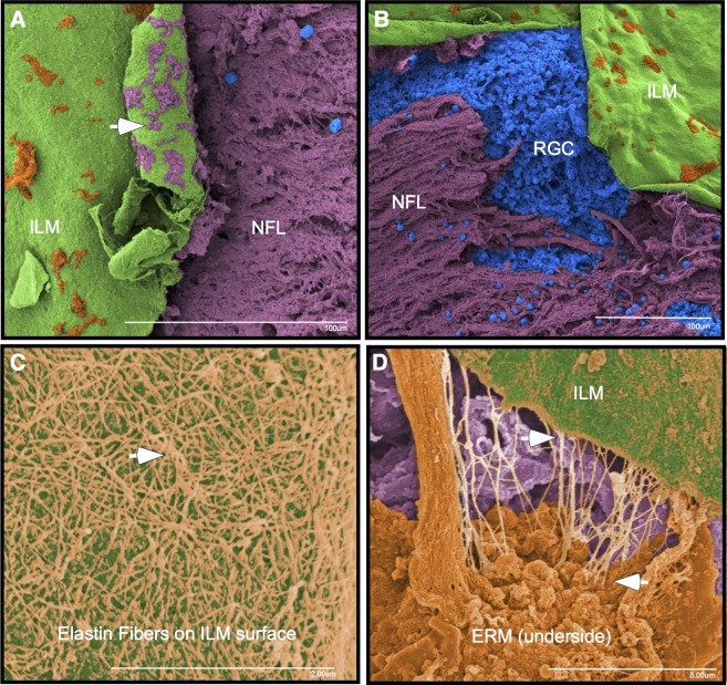
Purpose: The purpose of this study was to identify the structural and histological effects of a Tano diamond-dusted membrane scraper (DDMS) on the retinal surface after internal limiting membrane (ILM) abrasion in macular hole surgery.

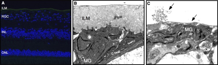
Methods: Institutional experimental study was performed in 11 eyes. All eyes underwent ILM abrasion in the operating room with a DDMS for macular hole repair as an alternative to traditional ILM peeling. Three human donor eyes underwent an identical procedure in the laboratory. Retinal tissues were removed by ILM abrasion with a DDMS during vitrectomy for macular hole repair and retinal tissues remaining in human donor eyes. Main outcome measures were microscopic and immunohistological characteristics of instrument tip tissues and retinal structure after ILM abrasion.

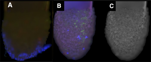
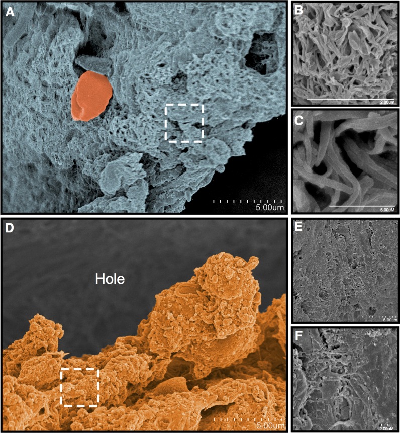
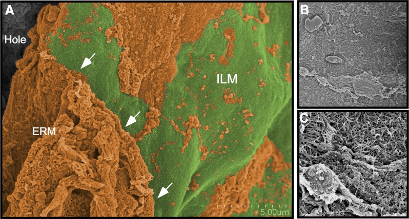
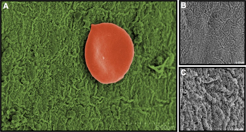
Results: The tips of the Tano DDMS showed evidence of cellular membranes and ILM removal. The retinas showed distinct areas of lamellar ILM removal without penetration of the retinal nerve fiber layer (RNFL).

Conclusions: Application of the Tano DDMS instrument is sufficient to remove membranes from the surface of the ILM and layers of the ILM without disruption of the underlying RNFL. Internal limiting membrane abrasion can be a useful and effective alternative to complete ILM removal for macular surgery.


Figures

Figure 1
Figure 1
Tano scraper tips show ILM fragments and cells. After they were used for treatment of patient retinas during macular hole surgery, the Tano scratchers were visualized with immunohistochemistry. (A) Numerous cells (blue) can be seen on sheets of tissue attached to the instrument tips. (B) Laminin A–positive tissue fragments (green) indicate ILM and cells (blue) on the Tano tips. (C) Control confocal fluorescence image of an unstained tip.
Figure 2
Figure 2
Tano scratcher applied to a macular hole in a postmortem eye. (A) Dissecting microscope image of a macular hole. The retina surrounding the macula was divided into four areas (numbered 14). (B) A low magnification scanning electron microscopy image of the specimen after treatment with a Tano scratcher. No Tano scratcher was applied to area 1. Light pressure was applied to area 2. Medium pressure was applied to area 3. Heavy pressure was applied to area 4, which shows disruption of the internal limiting membrane and exposure of the nerve fiber layer. Some of the cellular tissue has fallen into the hole.
Figure 3
Figure 3
Area 1: Macula without Tano scraper application. (AC) Away from the macular hole are vitreous collagenous elements on the surface of the retina. (DF) An epiretinal membrane (orange) is present at the edge of the macular hole, where there was no application of the Tano scraper. Higher magnification revealed cellular and collagenous elements, consistent with an epiretinal membrane.
Figure 4
Figure 4
Area 2: Macula with light Tano application. (A) After a light application of the Tano, an edge (arrows) of ILM (green) becomes apparent alongside a region where untreated ERM (orange) remains. (B, C) Higher magnification over the scraped area shows that some cell and tissue elements of the epiretinal membrane remain on the surface of the ILM.
Figure 5
Figure 5
Area 3: Macula with medium Tano application. (A) After a medium pressure scrape, nearly all cells and tissues from the epiretinal membrane were removed, revealing the ILM (green). (B, C) The ILM shows no overlying surface cells or tissues at higher magnification.
Figure 6
Figure 6
Area 4: Macula with heavy Tano application. (A, B) Heavy application of the Tano scraper caused a tear in the ILM (green) and subsequent exposure of the nerve fiber layer (purple) and cellular elements (blue). The underside of the ILM (arrow) shows fragments of the retinal nerve fiber layer (purple) remain attached to the ILM. (C) Fibers consistent with elastin (orange) were found between the ERM and ILM. (D) These fibers remain attached to the ILM and underside of the ERM.
Figure 7
Figure 7
Internal limiting membrane immunohistochemistry and transmission electron microscopy. (A) In human retinas treated with the DDMS, laminin A antibodies (green) identified the ILM. Cell nuclei were stained with DAPI (blue). ONL, outer nuclear layer. (B) Transmission electron microscopy image of untreated retinas showed Müller glial (MG) cell end feet processes at the edge of the ILM and filopodial insertions deeper within the ILM. (C) Treatment of retinas with medium pressure from the Tano resulted in removal of most of the ILM and the deepest Müller filopodial insertions, but preserved a thin layer of ILM over the MG end feet.

Similar articles

Cited by

References

    1. Gass JD.Idiopathic senile macular hole. Its early stages and pathogenesis. Arch Ophthalmol. 1988; 106: 629–639. - PubMed
    1. Gass JD.Risk of developing macular hole. Arch Ophthalmol. 1991; 109: 610–612. - PubMed
    1. Casuso LA,, Scott IU,, Flynn HW, Jr., et al. Long-term follow-up of unoperated macular holes. Ophthalmology. 2001; 108: 1150–1155. - PubMed
    1. Gupta P,, Yee KM,, Garcia P,, et al. Vitreoschisis in macular diseases. Br J Ophthalmol. 2011; 95: 376–380. - PMC - PubMed
    1. Kishi S,, Demaria C,, Shimizu K.Vitreous cortex remnants at the fovea after spontaneous vitreous detachment. Int Ophthalmol. 1986; 9: 253–260. - PubMed

Publication types

LinkOut - more resources