

Genetic dissection of horizontal cell inhibitory signaling in mice in complete darkness in vivo
- PMID: 26024096
- PMCID: PMC4451614
- DOI: 10.1167/iovs.15-16581
Genetic dissection of horizontal cell inhibitory signaling in mice in complete darkness in vivo
Abstract
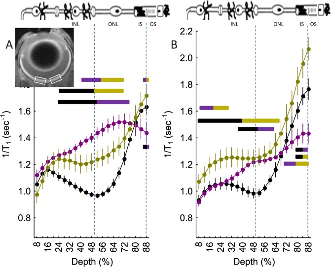
Purpose: To test the hypothesis that horizontal cell (HC) inhibitory signaling controls the degree to which rod cell membranes are depolarized as measured by the extent to which L-type calcium channels (LTCCs) are open in complete darkness in the mouse retina in vivo.
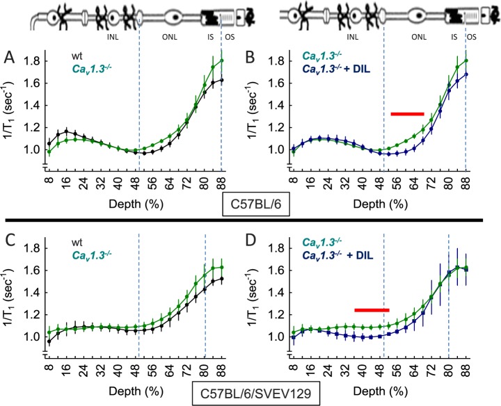
Methods: Dark-adapted wild-type (wt), CACNA1F (Ca(v)1.4(-/-)), arrestin-1 (Arr1(-/-)), and CACNA1D (Ca(v)1.3(-/-)) C57Bl/6 mice were studied. Manganese-enhanced MRI (MEMRI) evaluated the extent that rod LTCCs are open as an index of loss of HC inhibitory signaling. Subgroups were pretreated with D-cis-diltiazem (DIL) at a dose that specifically antagonizes Ca(v)1.2 channels in vivo.
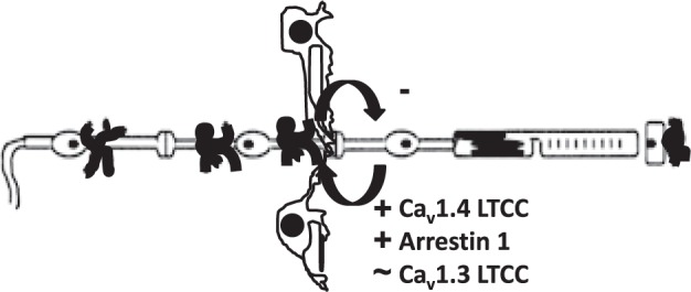
Results: Knockout mice predicted to have impaired HC inhibitory signaling (Ca(v)1.4(-/-) or Arr1(-/-)) exhibited greater than normal rod manganese uptake; inner retinal uptake was also supernormal. Genetically knocking out a closely associated gene not expected to impact HC inhibitory signaling (CACNA1D) did not generate this phenotype. The Arr1(-/-) mice exhibited the largest rod uptake of manganese. Manganese-enhanced MRI of DIL-treated Arr1(-/-) mice suggested a greater number of operant LTCC subtypes (i.e., Ca(v)1.2, 1.3, and 1.4) in rods and inner retina than that in DIL-treated Ca(v)1.4(-/-) mice (i.e., Ca(v)1.3). The Ca(v)1.3(-/-) + DIL-treated mice exhibited evidence for a compensatory contribution from Ca(v)1.2 LTCCs.
Conclusions: The data suggest that loss of HC inhibitory signaling is the proximate cause leading to maximally open LTCCs in rods, and possibly inner retinal cells, in mice in total darkness in vivo, regardless of compensatory changes in LTCC subtype manifested in the mutant mice.
Figures
Similar articles
-
D-cis-Diltiazem Can Produce Oxidative Stress in Healthy Depolarized Rods In Vivo.Invest Ophthalmol Vis Sci. 2018 Jun 1;59(7):2999-3010. doi: 10.1167/iovs.18-23829. Invest Ophthalmol Vis Sci. 2018. PMID: 30025125 Free PMC article.
-
Confirming a prediction of the calcium hypothesis of photoreceptor aging in mice.Neurobiol Aging. 2014 Aug;35(8):1883-91. doi: 10.1016/j.neurobiolaging.2014.02.020. Epub 2014 Mar 2. Neurobiol Aging. 2014. PMID: 24680323
-
Development of an MRI biomarker sensitive to tetrameric visual arrestin 1 and its reduction via light-evoked translocation in vivo.FASEB J. 2015 Feb;29(2):554-64. doi: 10.1096/fj.14-254953. Epub 2014 Oct 28. FASEB J. 2015. PMID: 25351983 Free PMC article.
-
Abundant L-type calcium channel Ca(v)1.3 (alpha1D) subunit mRNA is detected in rod photoreceptors of the mouse retina via in situ hybridization.Mol Vis. 2007 May 23;13:764-71. Mol Vis. 2007. PMID: 17563731 Free PMC article.
-
Calcium dynamics and regulation in horizontal cells of the vertebrate retina: lessons from teleosts.J Neurophysiol. 2017 Feb 1;117(2):523-536. doi: 10.1152/jn.00585.2016. Epub 2016 Nov 2. J Neurophysiol. 2017. PMID: 27832601 Free PMC article. Review.
Cited by
-
Melanopsin Phototransduction Contributes to Light-Evoked Choroidal Expansion and Rod L-Type Calcium Channel Function In Vivo.Invest Ophthalmol Vis Sci. 2016 Oct 1;57(13):5314-5319. doi: 10.1167/iovs.16-20186. Invest Ophthalmol Vis Sci. 2016. PMID: 27727394 Free PMC article.
-
Applications of Manganese-Enhanced Magnetic Resonance Imaging in Ophthalmology and Visual Neuroscience.Front Neural Circuits. 2019 May 14;13:35. doi: 10.3389/fncir.2019.00035. eCollection 2019. Front Neural Circuits. 2019. PMID: 31156399 Free PMC article. Review.
-
D-cis-Diltiazem Can Produce Oxidative Stress in Healthy Depolarized Rods In Vivo.Invest Ophthalmol Vis Sci. 2018 Jun 1;59(7):2999-3010. doi: 10.1167/iovs.18-23829. Invest Ophthalmol Vis Sci. 2018. PMID: 30025125 Free PMC article.
-
MRI of rod cell compartment-specific function in disease and treatment in vivo.Prog Retin Eye Res. 2016 Mar;51:90-106. doi: 10.1016/j.preteyeres.2015.09.001. Epub 2015 Sep 4. Prog Retin Eye Res. 2016. PMID: 26344734 Free PMC article. Review.
-
MRI of Retinal Free Radical Production With Laminar Resolution In Vivo.Invest Ophthalmol Vis Sci. 2016 Feb;57(2):577-85. doi: 10.1167/iovs.15-18972. Invest Ophthalmol Vis Sci. 2016. PMID: 26886890 Free PMC article.
References
-
- Carter-Dawson LD,, Lavail MM,, Sidman RL. Differential effect of the rd mutation on rods and cones in the mouse retina. Invest Ophthalmol Vis Sci. 1978; 17: 489–498. - PubMed
-
- Morgans CW,, Gaughwin P,, Maleszka R. Expression of the alpha1F calcium channel subunit by photoreceptors in the rat retina. Mol Vis. 2001; 7: 202–209. - PubMed
-
- Schmitz Y,, Witkovsky P. Dependence of photoreceptor glutamate release on a dihydropyridine-sensitive calcium channel. Neuroscience. 1997; 78: 1209–1216. - PubMed
-
- Baumann L,, Gerstner A,, Zong X,, Biel M,, Wahl-Schott C. Functional characterization of the L-type Ca2+ channel cav1.4alpha1 from mouse retina. Invest Ophthalmol Vis Sci. 2004; 45: 708–713. - PubMed
Publication types
MeSH terms
Substances
Grants and funding
LinkOut - more resources
Full Text Sources
Other Literature Sources
Molecular Biology Databases
