. 2015 May;56(5):3383-92.
doi: 10.1167/iovs.15-16606.

Hyperglycemia-suppressed expression of Serpine1 contributes to delayed epithelial wound healing in diabetic mouse corneas

Hyperglycemia-suppressed expression of Serpine1 contributes to delayed epithelial wound healing in diabetic mouse corneas

Haijing Sun et al. Invest Ophthalmol Vis Sci. 2015 May.

Abstract

Purpose: Patients with diabetes mellitus (DM) are at an increased risk for developing corneal complications, including delayed wound healing. The purpose of this study was to characterize the expression and the function of Serpine1 and other components of urokinase plasminogen activator (uPA)-proteolytic system in delayed epithelial wound healing in diabetic mouse corneas.

Methods: Mice of the strain C57BL/6 were induced to develop diabetes by streptozotocin, and wound-healing assays were performed 10 weeks afterward. Gene expression and/or distribution were assessed by real-time PCR, Western blotting, and/or immunohistochemistry. The role of Serpine1 in mediating epithelial wound closure was determined by subconjunctival injections of neutralizing antibodies in either normal or recombinant protein in diabetic corneas. Enzyme assay for matrix metalloproteinase (MMP)-3 was also performed.

Results: The expressions of Serpine1 (PAI-1), Plau (uPA), and Plaur (uPA receptor) were upregulated in response to wounding, and these upregulations were significantly suppressed by hyperglycemia. In healing epithelia, Plau and Serpine1 were abundantly expressed at the leading edge of the healing epithelia of normal and, to a lesser extent, diabetic corneas. Inhibition of Serpine1 delayed epithelial wound closure in normal corneas, whereas recombinant Serpine1 accelerated it in diabetic corneas. The Plau and MMP-3 mRNA levels and MMP-3 enzymatic activities were correlated to Serpine1 levels and/or the rates of epithelial wound closure.

Conclusions: Serpine1 plays a role in mediating epithelial wound healing and its impaired expression may contribute to delayed wound healing in DM corneas. Hence, modulating uPA proteolytic pathway may represent a new approach for treating diabetic keratopathy.


Figures
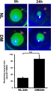
Figure 1
Figure 1
Delayed wound healing in STZ-induced type 1 diabetic mice. B6 mice (6 weeks old) were intraperitoneally injected with 50 mg/kg STZ daily for 5 days; control mice received citrate buffer (pH 4.5). Mice were tested for the levels of blood sugar at week 4 post injection. Mice with higher than 350 mg/dL blood glucose were used for wound-healing study at week 10 post STZ injection. A 1.5-mm wound was made in the center of the cornea and the healing process was visualized with fluorescein staining of the denuded area. Top: Representative images are shown. Bottom: The wound sizes were calculated by Adobe Photoshop (V6) Magic Wand Tool and Histogram (Adobe Systems, Inc., San Jose, CA, USA) and presented as the average of the number of pixels of the fluorescent-stained area. The error bars represent the SEM (n = 5) and indicated P values were generated using paired Student's t-test. Five independent experiments (n = 5 each condition) were performed.
Figure 2
Figure 2
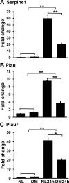
Real-time PCR verification of uPA system gene expression in healing versus homeostatic CECs of NL and DM mice ([A] Serpine1, [B] Plau, [C] Plaur). Corneal epithelial cells were collected from nondiabetic (NL) and STZ diabetic (DM) mouse corneas during epithelium-debridement or from the wound bed 24 hours after wounding (24h) (Fig. 1) and were subjected to real-time PCR analysis. The fold increase over that of the naïve corneas (value 1) is the mean ± SD of three samples (n = 3) for each condition with β-actin as the internal control for normalization. The results are representative of two independent experiments. *P < 0.05; **P < 0.01 (one-way ANOVA).
Figure 3
Figure 3
Western blotting analysis of Serpine1 and Plau expression in healing versus homeostatic CECs of NL and DM mice. The scraped CECs from NL and DM mouse corneas for creating a wound and from the wound bed 24 hours after wounding, as described in Figure 2, were processed for Western blotting with β-actin as internal control for protein loading. (A) Two samples for each condition were run on the same gel. After transfer, the membrane was probed first with Serpine1 (48 kDa), after stripping, with Plau (34 kDa), and then actin (42 kDa). (B) The band intensities were calculated after background-subtracting and normalized with actin intensity using Carestream Molecular Imaging Software. The fold increase over that of the normal, unwounded cornea (value 1) is the mean ± SD of three samples (n = 2, two corneas each) for each condition with β-actin as the internal control for normalization. The results are representative of two independent experiments (two each). *P < 0.05 (one-way ANOVA).
Figure 4
Figure 4
Serpine1 and Plau expression and distribution in wound-healing corneas of NL and DM mice assessing by immunohistochemistry. B6 and STZ B6 mouse corneas were wounded as described in Figure 1. At 24 hpw, corneas with remaining wounds were excised and snap-frozen in OCT compound, followed by sectioning and immunostaining with antibodies against Serpine1 or Plau. Unwounded corneas of NL and DM mice also were stained; DAPI was used to stain nuclei. (A) The photographs were taken at the center of unwounded cornea or the leading edge of healing corneas with the same exposure time and other settings. (B) The fluorescence intensity was semiquantitatively analyzed using ImageJ. The fold increase over that of the normal, unwounded cornea (value 1) is the mean ± SD of three samples (n = 3) for each condition. The results are representative of two independent experiments. *P < 0.05; **P < 0.01 (one-way ANOVA).
Figure 5
Figure 5
Effects of Serpine1 on corneal epithelial wound healing in NL and DM mice. Serpine1 neutralizing antibody (2.5 μg/5 μL), with the same dosage of rabbit IgG as the control, were injected into subconjunctival space of NL and STZ B6 mice or 500 ng/5 μL recombinant Serpine1 (Millipore, Billerica, MA, USA) to STZ B6 mice 4 hours before wounding. A 1.5-mm wound was allowed to heal for 24 hours, fluorescence stained, and photographed (A). These figures are representative of six corneas per condition from two independent experiments. (B) The wound sizes were presented as the average number of pixels of the fluorescent-stained area (Fig. 1). The error bars represent the SEM (n = 6) and indicated P values were generated using paired Student's t-test. Two independent experiments were performed.
Figure 6
Figure 6
Effects of Serpine1 levels on Plau and MMP-3 expressions in healing versus homeostatic CECs of NL and DM mice. Corneal endothelial cells were scraped off from NL and DM mouse corneas for creating a wound (0 hour) from the wound bed 24 hpw as described in Figure 5. The collected CECs were subjected to real-time PCR. The data were presented as mean fold change in the expression of indicated gene over nondiabetic, 0-hour CECs. The error bars represent the SEM (n = 3) and indicated P values were generated using paired Student's t-test. Two independent experiments were performed.
Figure 7
Figure 7
Effects of Serpine1 levels on MMP-3 enzymatic activity in healing versus homeostatic CECs of NL and DM mice. Corneal endothelial cells were scraped off from NL and DM mouse corneas for creating a wound (0 hour) and from the wound bed 24 hpw as described in Figure 5. These collected CECs were lysed in RIPA buffer and the cell extracts after debris removal were analyzed by MMP-3 Activity kit from Abcam using 50 μg total proteins from each sample. The data were presented as mean fold change in MMP-3 enzymatic activities over nondiabetic, 0-hour CECs as 1. The error bars represent the SEM (n = 3) and indicated P values were generated using paired Student's t-test. Two independent experiments were performed.

Similar articles

Cited by

References

    1. Clark EA,, Grabstein KH,, Gown AM,, et al. Activation of B lymphocyte maturation by a human follicular dendritic cell line, FDC-1. J Immunol. 1995; 155: 545–555. - PubMed
    1. Threatt J,, Williamson JF,, Huynh K,, Davis RM. Ocular disease, knowledge and technology applications in patients with diabetes. Am J Med Sci. 2013; 345: 266–270. - PMC - PubMed
    1. Kaji Y. Prevention of diabetic keratopathy. Br J Ophthalmol. 2005; 89: 254–255. - PMC - PubMed
    1. Pflugfelder SC. Is autologous serum a tonic for the ailing corneal epithelium? Am J Ophthalmol. 2006; 142: 316–317. - PubMed
    1. Bikbova G,, Oshitari T,, Tawada A,, Yamamoto S. Corneal changes in diabetes mellitus. Curr Diabetes Rev. 2012; 8: 294–302. - PubMed

Publication types

MeSH terms