. 2015 May 29;10(5):e0126817.
doi: 10.1371/journal.pone.0126817. eCollection 2015.

A Method for 3D Histopathology Reconstruction Supporting Mouse Microvasculature Analysis

Affiliations

A Method for 3D Histopathology Reconstruction Supporting Mouse Microvasculature Analysis

Yiwen Xu et al. PLoS One. .

Abstract

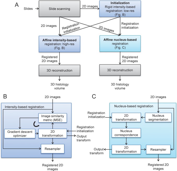
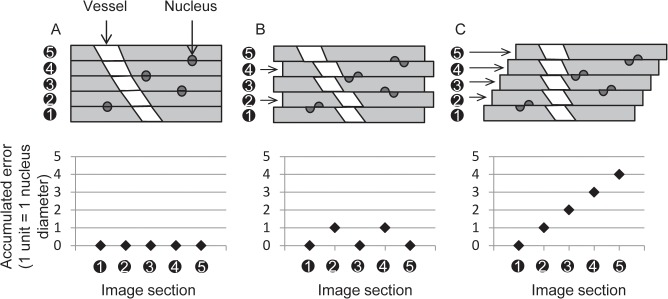
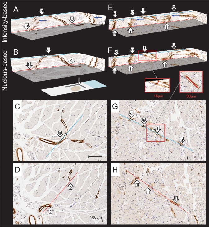
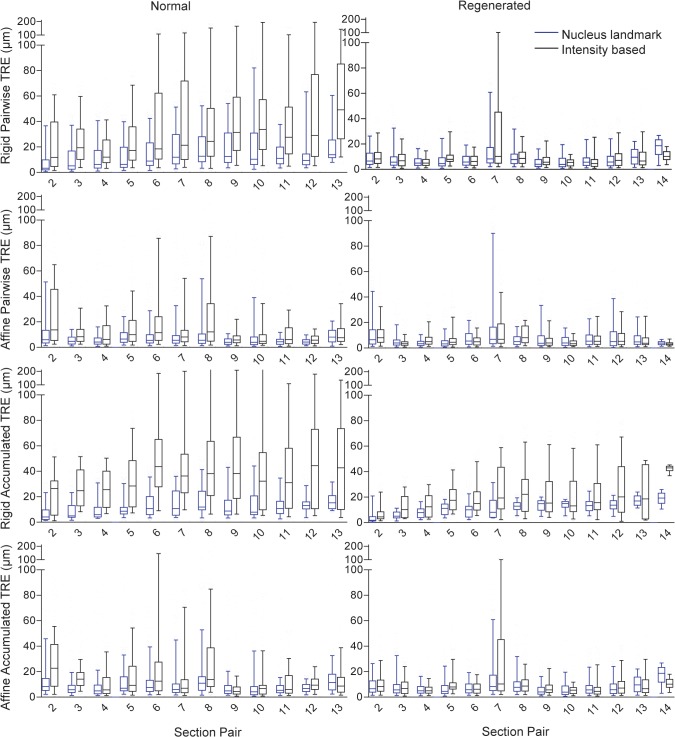
Structural abnormalities of the microvasculature can impair perfusion and function. Conventional histology provides good spatial resolution with which to evaluate the microvascular structure but affords no 3-dimensional information; this limitation could lead to misinterpretations of the complex microvessel network in health and disease. The objective of this study was to develop and evaluate an accurate, fully automated 3D histology reconstruction method to visualize the arterioles and venules within the mouse hind-limb. Sections of the tibialis anterior muscle from C57BL/J6 mice (both normal and subjected to femoral artery excision) were reconstructed using pairwise rigid and affine registrations of 5 µm-thick, paraffin-embedded serial sections digitized at 0.25 µm/pixel. Low-resolution intensity-based rigid registration was used to initialize the nucleus landmark-based registration, and conventional high-resolution intensity-based registration method. The affine nucleus landmark-based registration was developed in this work and was compared to the conventional affine high-resolution intensity-based registration method. Target registration errors were measured between adjacent tissue sections (pairwise error), as well as with respect to a 3D reference reconstruction (accumulated error, to capture propagation of error through the stack of sections). Accumulated error measures were lower (p < 0.01) for the nucleus landmark technique and superior vasculature continuity was observed. These findings indicate that registration based on automatic extraction and correspondence of small, homologous landmarks may support accurate 3D histology reconstruction. This technique avoids the otherwise problematic "banana-into-cylinder" effect observed using conventional methods that optimize the pairwise alignment of salient structures, forcing them to be section-orthogonal. This approach will provide a valuable tool for high-accuracy 3D histology tissue reconstructions for analysis of diseased microvasculature.


Conflict of interest statement

Competing Interests: The authors have declared that no competing interests exist.

Figures

Fig 1
Fig 1. Diagram depicting the registration methods.
The flow diagram in (A) depicts the overall experimental process, with (B) and (C) giving exploded views of the intensity-based and nucleus-based registration steps. Both approaches were initialized with a rigid, low-resolution intensity-based registration. The intensity-based registrations (B) were done using a standard iterative optimization loop. The nucleus-based registration (C) was computed non-iteratively in closed form based on automatically segmented and corresponded nucleus landmarks. Both methods were executed pairwise on each adjacent section pair, and as a final step these pairwise registrations were composed to form the final 3D reconstructed volume.
Fig 2
Fig 2. Comparison of the alignment of bisected nuclei when measuring the accumulated registration error.
(A) The ideal error-free reference reconstruction, with bisected nuclei aligned with minimum residual error (a pairwise target registration error between corresponding halves of bisected nuclei of zero is depicted). (B) A reconstruction aligning nuclei with spatially unbiased error; but vessel connectedness (topology) and angle (geometry) are mostly conserved. (C) A reconstruction optimizing pairwise alignment of salient structures (the vessel cross sections in this example) preserves vessel topology but not geometry. Note that the pairwise target registration errors in (B) and (C) are similar, despite the lack of geometry preservation in C. The accumulated target registration error does capture the difference between (B) and (C); the plots in the bottom row indicate increasing accumulated error through the stack of sections.
Fig 3
Fig 3. Nucleus correspondence method.
An illustration depicting the approach to establishing correspondence of nucleus p in section I with its best matching nucleus in adjacent section J. In this example, the candidate nuclei on section J are p’, q 1, and q 2, lying within a dashed circle of radius T centred on p (only 3 of the 18 candidate nuclei within the circle are illustrated here for simplicity). The candidate nucleus with the most similar surrounding tissue appearance is selected to correspond to p. Surrounding tissue appearance similarity is measured using the MSE image similarity metric, comparing the local square region I(p) centered on p with the local square regions J(p’), J(q 1 ), and J(q 2 ) centered on the candidates p’, q1, and q2. In this example, since MSE(I(p), J(p’)) < MSE(I(p), J(q1)) and MSE(I(p), J(p’)) < MSE(I(p), J(q2)), p is corresponded with p’.
Fig 4
Fig 4. 3D and 2D histology comparisons.
2D histology sections (pixel size 0.25 μm × 0.25 μm) and corresponding 3D reconstruction (voxel size of 0.25 μm × 0.25 μm × 5 μm) of serial histology sections of a normal (A-D) and regenerated mouse (E-H) TA post-femoral artery excision, immunostained for smooth muscle alpha-actin. A and E are registered using affine intensity based registration. B and F are registered using affine nucleus based registration. Within each column, the dashed lines indicate correspondence (according to color) between parts of the 2D sections and their locations on the 3D views. Also within each column, the lower case letter labels indicate correspondence between vessel cross sections on the 2D sections and their homologous locations within the 3D views. Blue arrows indicate incorrect vessel wall discontinuities arising from reconstruction error. The insets in the red boxes show 2D and 3D diameter measurements of the same vessel; note that the 2D measurement overestimates the 3D measurement by a factor of >6. Scale bars 100 μm.
Fig 5
Fig 5. Registration accuracy measurement values.
Box plots of the rigid and affine target registration error (TRE) computed for each adjacent pair of sections (pairwise) and propagated throughout the 3D reconstruction (accumulated).

References

    1. Pietra GG, Capron F, Stewart S, Leone O, Humbert M, Robbins IM, et al. Pathologic assessment of vasculopathies in pulmonary hypertension. J Am Coll Cardiol. 2004;43(12 Suppl S):25S–32S. Epub 2004/06/15. 10.1016/j.jacc.2004.02.033 . - DOI - PubMed
    1. Csernus B, Ficsor L, Molnar B, Sapi Z. Comparison of the vasculature of myxofibrosarcoma and myxoid liposarcoma using 3d histological reconstruction. Histopathology. 2008;53(Journal Article):410–1.
    1. Cifor A, Bai L, Pitiot A. Smoothness-guided 3-D reconstruction of 2-D histological images. NeuroImage. 2011;56(1):197–211. 10.1016/j.neuroimage.2011.01.060 - DOI - PubMed
    1. Schwier M, Bohler T, Hahn HK, Dahmen U, Dirsch O. Registration of histological whole slide images guided by vessel structures. Journal of pathology informatics. 2013;4(Suppl):S10–3539.109868. Print 2013. 10.4103/2153-3539.109868 - DOI - PMC - PubMed
    1. Walker EJ, Shen F, Young WL, Su H. Cerebrovascular Casting of the Adult Mouse for 3D Imaging and Morphological Analysis. J Vis Exp. 2011;(57). Epub 2012/03/23. 10.3791/2958 . - DOI - PubMed

Publication types

LinkOut - more resources