. 2015 May 29;10(5):e0127712.
doi: 10.1371/journal.pone.0127712. eCollection 2015.

TMEM120A and B: Nuclear Envelope Transmembrane Proteins Important for Adipocyte Differentiation

Affiliations

TMEM120A and B: Nuclear Envelope Transmembrane Proteins Important for Adipocyte Differentiation

Dzmitry G Batrakou et al. PLoS One. .

Abstract

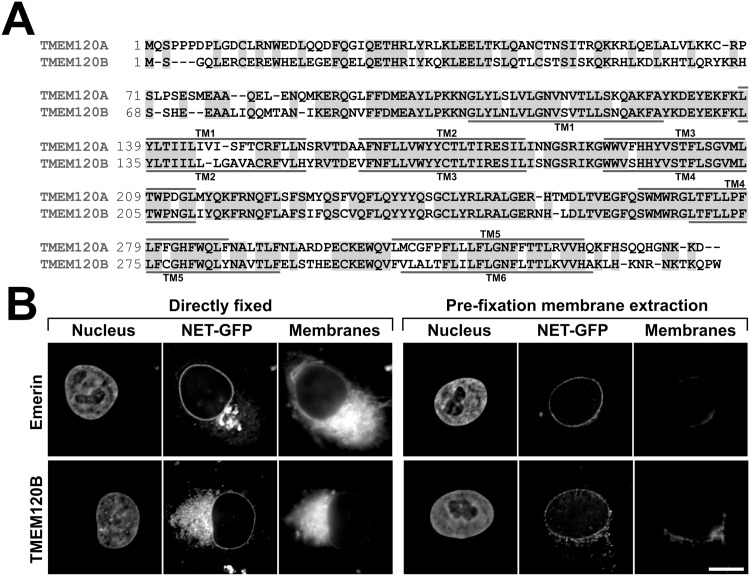
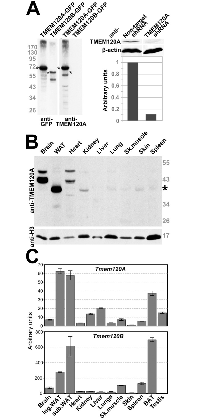
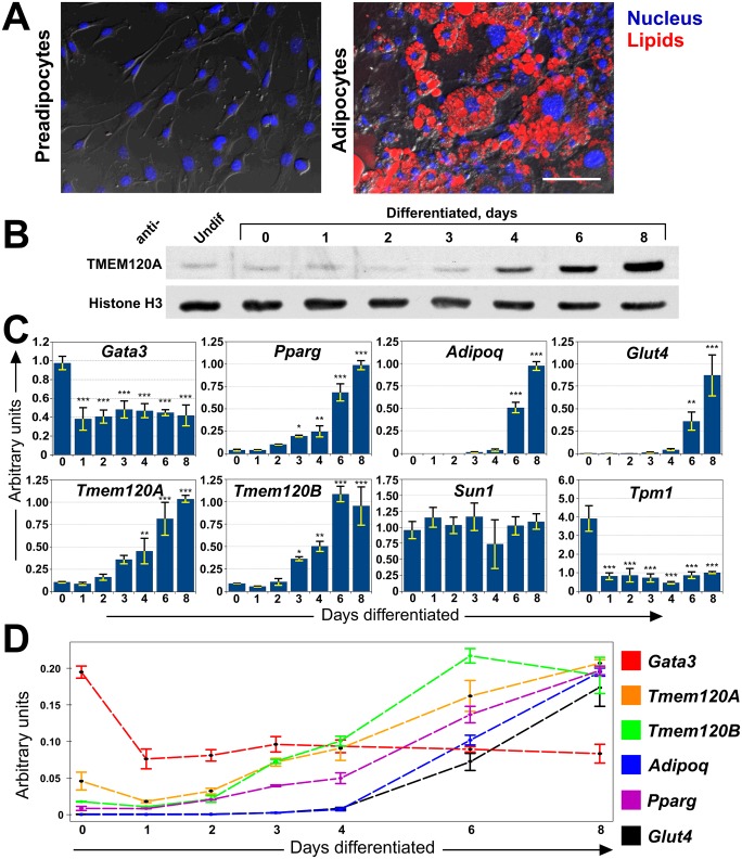
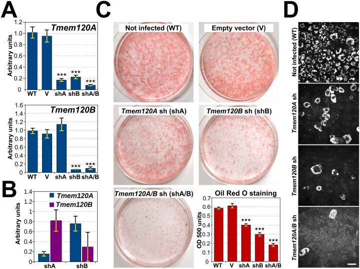
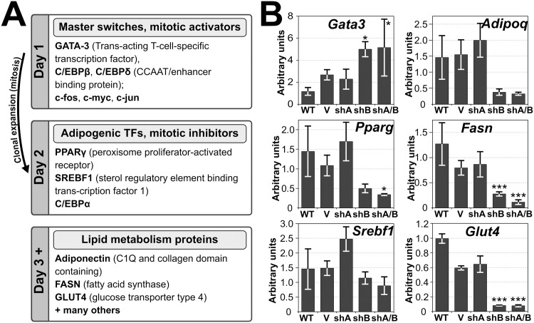
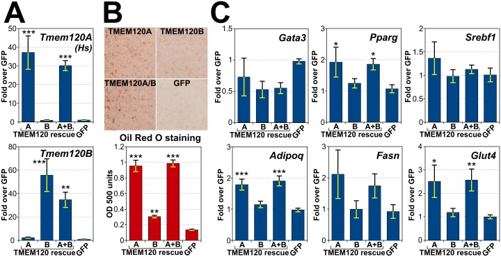
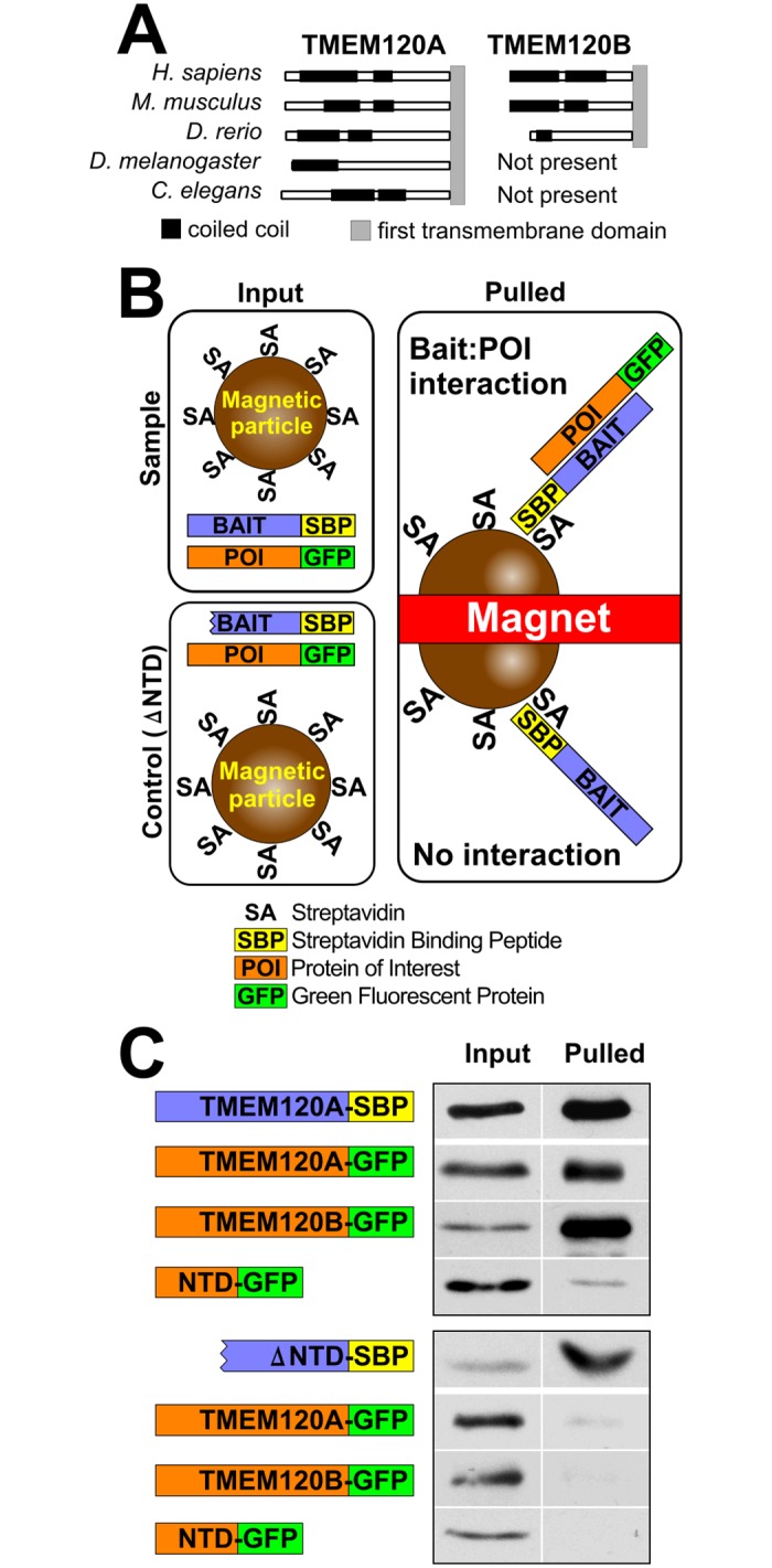
Recent work indicates that the nuclear envelope is a major signaling node for the cell that can influence tissue differentiation processes. Here we present two nuclear envelope trans-membrane proteins TMEM120A and TMEM120B that are paralogs encoded by the Tmem120A and Tmem120B genes. The TMEM120 proteins are expressed preferentially in fat and both are induced during 3T3-L1 adipocyte differentiation. Knockdown of one or the other protein altered expression of several genes required for adipocyte differentiation, Gata3, Fasn, Glut4, while knockdown of both together additionally affected Pparg and Adipoq. The double knockdown also increased the strength of effects, reducing for example Glut4 levels by 95% compared to control 3T3-L1 cells upon pharmacologically induced differentiation. Accordingly, TMEM120A and B knockdown individually and together impacted on adipocyte differentiation/metabolism as measured by lipid accumulation through binding of Oil Red O and coherent anti-Stokes Raman scattering microscopy (CARS). The nuclear envelope is linked to several lipodystrophies through mutations in lamin A; however, lamin A is widely expressed. Thus it is possible that the TMEM120A and B fat-specific nuclear envelope transmembrane proteins may play a contributory role in the tissue-specific pathology of this disorder or in the wider problem of obesity.


Conflict of interest statement

Competing Interests: The authors have declared that no competing interests exist.

Figures

Fig 1
Fig 1. Paralog TMEM120B and its targeting to the NE.
(A) Sequence alignment between mouse TMEM120A and TMEM120B. Identical residues are highlighted with gray shading and predicted transmembrane spans (TM) are indicated. (B) Targeting of TMEM120B to the NE. Left panels, directly fixed HT1080 cells expressing TMEM120B fused to GFP or positive control emerin-GFP (NET-GFP) stained for DNA (Nucleus) or with the membrane dye DiOC6 (Membranes). Right panels, equivalent cells were extracted with 0.2% Triton X-100 prior to fixation. This treatment should remove membranes and proteins not strongly bound to structures such as the nucleo/cytoskeleton and chromatin. Scale bar, 10 μm.
Fig 2
Fig 2. Preferential expression of TMEM120A and B in fat.
(A) TMEM120A-specific antibodies. Antibodies generated in rabbit to amino acid residues 52–71 were tested against lysates from HT1080 cells expressing TMEM120A and B as GFP fusions and 6 days differentiated 3T3-L1 stably expressing either non-target shRNA or TMEM120A-specific shRNA. The left panel is HT1080 cells exogenously expressing TMEM120-GFP fusions reacted either with the GFP antibody or with the TMEM120A antibody; asterisks denote TMEM120A and B GFP fusion proteins. The right panel, top, is quantitative Western blot of 3T3-L1 cell lysates from the shRNA knockdown and controls reacted with either the anti-TMEM120A or anti-β-actin antibody; bottom, quantification of the TMEM120A signal from the above blot, normalized to β-actin. (B) Lysates were generated from freshly isolated mouse tissue and reacted on Western with the TMEM120A antibodies. Of the tissues tested, WAT expressed by far the strongest a band for the expected size of TMEM120A; however, two bands larger than the size for any predicted splice variant were also strongly expressed in brain and to a lesser degree in heart. Thus it is unclear if these are TMEM120A variants. (C) Total RNA was extracted from mouse tissues and subjected to qRT-PCR using specific primers for Tmem120A and Tmem120B. The data is expressed as the fold-increase over the lowest level (skin for Tmem120A and testis for Tmem120B). Error bars indicate the standard deviation for three technical repeats.
Fig 3
Fig 3. Induction of TMEM120A, TMEM120B and adipogenic markers during adipogenesis.
(A) The 3T3-L1 in vitro differentiation system. Left panel, 3T3-L1 pre-adipocytes have a normal fibroblast morphology and do not stain with LipidTOX which stains lipid droplets. Right panel, after induction by treatment with iso-butyl methyl xanthine, dexamethasone and insulin the cells begin to accumulate lipid droplets by day 4 and this increases so that by day 8 (shown) the vast majority of cells have generated many large lipid droplets that stain with LipidTOX. Scale bar, 100 μm. (B) Lysates were generated from cells over a timecourse of 3T3-L1 induction and reacted with Tmem120A antibodies or with histone H3 antibodies as a loading control. The protein is induced over time, with the major accumulation between days 3 and 8. (C) RNA was extracted from a timecourse of 3T3-L1 adipogenesis and the relative mRNA levels for Tmem120A, Tmem120B, Gata3, Pparg, AdipoQ, Glut4 and controls Sun1 and Tpm1 measured by qRT-PCR. Asterisks indicate statistical significance (* p<0.05, ** p<0.01, *** p<0.001). (D) A subset of the same data overlaid as line plots for direct comparison clearly shows the earlier activated Gata3 dropping prior to activation of the Tmem120A and B genes which become activated in relative levels at a faster rate than the master regulator Pparg.
Fig 4
Fig 4. Both TMEM120 proteins are critical for adipogenesis.
(A) TMEM120A and TMEM120B were knocked down in 3T3-L1 cells by viral transduction of shRNA constructs both individually and together and the cells were induced pharmacologically to differentiate. RNA was extracted on day 8 and the relative levels of transcripts from both genes using Sun1 as a control was determined by qRT-PCR. Notably TMEM120B knockdown affected both TMEM120B and TMEM120A levels in the cells induced for differentiation. (B) TMEM120B knockdown does not affect the minimal TMEM120A levels in non-differentiated cells. The values plotted represent the ratios of Tmem120 levels in knockdown cell lines to corresponding levels in cells transduced with empty vector. (C) To follow adipocyte differentiation the cells were stained with Oil Red O, a dye that intercalates in lipid droplets (images from day 12 of differentiation). To quantify the extent of differentiation by this measure, the Oil Red O dye was extracted from the cells with isopropanol and measured in a spectrophotometer (lower right panel, timepoint taken at 8 days differentiation). Both TMEM120 proteins knockdowns reduced lipid droplet accumulation by this measure significantly on their own and both together reduced lipid droplets by roughly 70%. (D) Lipid droplet accumulation was also assessed by CARS microscopy, a method based on Raman spectroscopy that directly highlights these structures without the use of dyes (images taken at 8 days differentiation). Scale bar, 50 μm.
Fig 5
Fig 5. Both early expressed adipogenic transcription factors and late expressed lipid metabolism proteins transcripts are targeted by TMEM120 protein functions.
(A) Well-defined protein functional cascades during pharmacologically induced 3T3-L1 differentiation. Early upregulated or downregulated genes include Gata3, Srebf1 and master regulator of adipogenesis, Pparg. Later in differentiation, levels of their targets, proteins involved in lipid methabolism, raise. [44,45] (B) RNA extracted from the TMEM120A and TMEM120B knockdown cells at day 8 of the adipogenic differentiation in Fig 4 were analyzed by qRT-PCR for effects on genes known to be important for adipogenesis (e.g. Gata3, Pparg, Srebf1) or fat metabolism (e.g. Fasn, AdipoQ, Glut4). Both NETs had effects, but the inhibitory effect of TMEM120B knockdown was stronger for most of the genes tested.
Fig 6
Fig 6. Reintroduction of TMEM120 proteins rescues adipogenesis.
Either shRNA resistant TMEM120A, TMEM120B, the combination of the two, or a GFP control were expressed in Tmem120A/B double knockdown cells and the cells were induced pharmacologically to differentiate. (A) Levels of human Tmem120A and mouse Tmem120B transcripts were assessed in the rescue cell lines from 8 days post adipogenic induction. (B) Oil Red O staining of the rescue cell lines. Top panel, cells stained with Oil Red O. Bottom panel, quantification by spectrophotometry at OD 500 of the dye extracted from cells. (C) RNA extracted from cells in this experiment was used to measure transcript levels of important adipogenic transcription factors and lipid metabolism proteins.
Fig 7
Fig 7. Oligomerization of TMEM120A and B.
(A) Coiled-coil domains are strongly predicted within the N-terminal regions of both TMEM120A and B using the COILS2 server for all organisms having both TMEM120 genes and predicted in the single protein for organisms having only one TMEM120 gene. (B) Schematic of assay and expected outcomes. Cells co-express streptavidin binding protein (SBP) fused to one TMEM120 protein and GFP fused to the same or another TMEM120 protein. The cells are lysed and the SBP fusion proteins are pulled from the lysate with streptavidin conjugated to magnetic beads. If the other TMEM120 protein fused to GFP is pulled out together with the SBP tagged protein it indicates interaction between the two TMEM120 proteins expressed. As a negative sontrol, TMEM120A-SBP fusion lacking predicted coil-coiled domains (deleted 2–105 amino acids, ΔNTD) was used. (C) Proteins pulled out of the lysates by the magnetic streptavidin coated beads were analyzed by Western blot using antibodies to GFP to detect the GFP fusions and streptavidin to test the SBP fusions. This confirmed both hetero- and homo-dimer formation are possible among TMEM120 proteins. The soluble N-terminal fragment containing the predicted coiled coils also interacted with the full-length protein, but not the ΔNTD mutant.

Similar articles

Cited by

References

    1. WHO. Obesity and overweight [Internet]. 2011. Available: http://www.who.int.mediacentre/factsheets/fs311/en/
    1. Fall T, Ingelsson E. Genome-wide association studies of obesity and metabolic syndrome. Mol Cell Endocrinol. 2014;382: 740–757. 10.1016/j.mce.2012.08.018 - DOI - PubMed
    1. Willyard C. Heritability: The family roots of obesity. Nature. 2014;508: S58–60. 10.1038/508S58a - DOI - PubMed
    1. Ali AT, Hochfeld WE, Myburgh R, Pepper MS. Adipocyte and adipogenesis. Eur J Cell Biol. Elsevier GmbH.; 2013;92: 229–236. 10.1016/j.ejcb.2013.06.001 - DOI - PubMed
    1. De Las Heras JI, Meinke P, Batrakou DG, Srsen V, Zuleger N, Kerr AR, et al. Tissue specificity in the nuclear envelope supports its functional complexity. Nucleus. 2013/11/12 ed. 2013;4: 460–477. doi:26872 [pii] 10.4161/nucl.26872 - DOI - PMC - PubMed

Publication types

MeSH terms