. 2015 May 29:5:10627.
doi: 10.1038/srep10627.

A novel inhibitory mechanism of MRTF-A/B on the ICAM-1 gene expression in vascular endothelial cells

Affiliations

A novel inhibitory mechanism of MRTF-A/B on the ICAM-1 gene expression in vascular endothelial cells

Ken'ichiro Hayashi et al. Sci Rep. .

Abstract

The roles of myocardin-related transcription factor A (MRTF-A) and MRTF-B in vascular endothelial cells are not completely understood. Here, we found a novel regulatory mechanism for MRTF-A/B function. MRTF-A/B tend to accumulate in the nucleus in arterial endothelial cells in vivo and human aortic endothelial cells (HAoECs) in vitro. In HAoECs, nuclear localization of MRTF-A/B was not significantly affected by Y27632 or latrunculin B, primarily due to the reduced binding of MRTF-A/B to G-actin and in part, to the low level of MRTF-A phosphorylation by ERK. MRTF-A/B downregulation by serum depletion or transfection of siRNA against MRTF-A and/or MRTF-B induced ICAM-1 expression in HAoECs. It is known that nuclear import of nuclear factor-κB (NF-κB) plays a key role in ICAM-1 gene transcription. However, nuclear accumulation of NF-κB p65 was not observed in MRTF-A/B-depleted HAoECs. Our present findings suggest that MRTF-A/B inhibit ICAM-1 mRNA expression by forming a complex with NF-κB p65 in the nucleus. Conversely, downregulation of MRTF-A/B alleviates this negative regulation without further translocation of NF-κB p65 into the nucleus. These results reveal the novel roles of MRTF-A/B in the homeostasis of vascular endothelium.


Figures

Figure 1
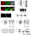
Figure 1. Subcellular localization of MRTF-A/B in human arterial endothelial cells.
(a) Immunohistochemical findings of MRTF-A and MRTF-B in the endothelium of normal human renal interlobular artery. Paraffin-embedded serial transverse sections were stained with HE or an antibody against CD34, MRTF-A or MRTF-B. Representative images are shown. (b and c) HAoECs were first cultured in HEC-C1 medium. For the last 24 h, they were cultured in either HEC-C1 medium or 0.1 × HEC-C1 medium. Statistical differences were calculated using student’s t-test. (d) NIH3T3 cells were cultured in Dulbecco’s modified Eagle’s medium supplemented with 10% fetal calf serum and in HEC-C1 medium for the last 24 h. Cells were stained with anti-MRTF-A antibody or anti-MRTF-B antibody (red) and Hoechst 33258 (blue). Representative images from at least three independent experiments are shown. Images were quantified as described in Materials and Methods: nuclear-specific localization (N), diffuse distribution in the nucleus and the cytoplasm (NC), and cytoplasmic localization (C) (left column).
Figure 2
Figure 2. Effects of Y27632 on the subcellular localization of MRTF-A/B in HAoECs.
HAoECs were cultured in HEC-C1 medium. For the last 12 h, they were treated with vehicle or 10 μM Y27632. (a) Whole cell lysates were subjected to IB analyses with the indicated antibodies. α↕tubulin was used as a loading control. (b and c) Cells were stained either with anti-MRTF-A antibody or anti-MRTF-B antibody (green), phalloidine-Alexa 568 (red), and Hoechst 33258 (blue). Representative images from at least three independent experiments are shown. Images were quantified as described in the legend for Fig. 1.
Figure 3
Figure 3. Properties of MRTF-A/B in HAoECs and HaCaT cells.
HaCaT cells were cultured in Dulbecco’s modified Eagle’s medium supplemented with 10% fetal calf serum and in HEC-C1 medium for the last 24 h. HAoECs were cultured in HEC-C1 medium. (a) HaCaT cells were stained with anti-MRTF-A antibody or anti-MRTF-B antibody (red), phalloidine-Alexa 488 (green), and Hoechst 33258 (blue). Representative images from at least three independent experiments are shown. (b) IB analysis shows the expression levels of MRTF-A/B and proteins involved in their nuclear import and export in HAoECs and HaCaT cells (upper panel). Whole cell lysates (WL) were subjected to IB with the indicated antibodies. α□tubulin was used as a loading control. RT-PCR analyses for monitoring the expression of MRTF-A isoforms (MAL fl [fl], variant X1 [X1], and variant X2 [X2]) in HAoECs and HaCaT cells (lower panel). PCR products were sampled at the indicated time points after 20 to 27 cycles and separated on 1.2% agarose gels. (c) Nuclear accumulation of exogenously expressed MRTF-A in HAoECs. HAoECs expressing Flag-tagged mouse MRTF-A (MAL fl) were stained with anti-Flag antibody (red) and Hoechst 33258 (blue). (d and e) IB analysis shows the expression levels of MRTF-A/B and β−actin in the whole cell extracts (WE) from HAoECs and HaCaT cells (d). The respective WE were subjected to IP analyses with a control antibody (cntl-Ab) or either anti-MRTF-A antibody (e left panel) or anti-MRTF-B antibody (e right panel). The IP/IB analyses were performed with the indicated antibodies. Positions of molecular weight markers (kDa) are indicated between the IB panels. (f) Actin fractionation. HAoECs and HaCaT cells were either left untreated (jasp-) or treated with jasplakinolide (0.3 μM; jasp+) for the last 60 min. The respective lysates (L) were separated into supernatant (S) and pellet (P) fractions by centrifugation, and they were subjected to IB with anti-β−actin antibody. (g) ERK phosphorylation of MRTF-A. WL from the indicated cells were subjected to IB with anti-MRTF-A or the phospho-specific MRTF-A antibody (left panel). WE from respective cells expressing Flag-tagged mouse MRTF-A (MAL fl) were subjected to IP/IB analysis with the indicated antibodies (right panel).
Figure 4
Figure 4. Relation between ERK activation and subcellular localization of MRTF-A in HAoECs and HaCaT cells.
(a,b, and c) HAoECs and HACaT cells were cultured in 0.1× HEC-C1 medium for 24 h, and then they were restimulated with HEC-C1 medium for 30 min and overnight (ON). Whole cell lysates at the indicated time points were subjected to IB with the indicated antibodies (a). HAoECs (b) and HaCaT cells (c) were stained with anti-ERK antibody (green) and anti-p-ERK antibody (red). Representative images from at least three independent experiments are shown. (d and e) Subcellular localization of exogenously expressed wild-type (wt) and mutant MRTF-As in HAoECs and HaCaT cells. HAoECs (d) and HaCaT cells (e) were transfected with each of the indicated Flag-tagged MRTF-A expression plasmids and were cultured in HEC-C1 medium containing either vehicle or 10 μM U0126 for 24 h. Cells were stained with anti-Flag antibody (red) and Hoechst 33258 (blue). Representative images from at least three independent experiments are shown. Images were quantified as described in the legend for Fig. 1. Statistical differences were calculated using student’s t-test. *P = 8.2580 × 10−5, **P = 0.0002, #P = 8.4164 × 10−7, and ##P = 1.5908 × 10−5 versus the values of MRTF-A wt in the respective localization categories (e).
Figure 5
Figure 5. Upregulation of ICAM-1 expression in MRTF-A/B-depleted HAoECs.
(a) Expression levels of MRTF-A/B, ICAM-1, endothelial cell markers, and transcription factors. HAoECs were transfected with control (cntl) siRNA or either anti-MRTF-A siRNA or anti-MRTF-B siRNA and were cultured for 2 days in HEC-C1 medium. For the last 24 h, they were cultured in HEC-C1 medium (lane 1) or in 0.1× HEC-C1 medium (lanes 2–4). Whole cell lysates (WL) were subjected to IB with the indicated antibodies. α−tubulin and GAPDH were used as loading controls. (b) Quantitative real-time RT-PCR analyses for MRTF-A/B and ICAM-1 mRNA expressions in HAoECs. Expression levels of the indicated mRNAs were normalized to GAPDH mRNA. The levels of the respective mRNAs in cntl siRNA-transfected HAoECs cultured in HEC-C1 medium were set at 100% (means ± SEMs of three independent experiments). (c) HAoECs were transfected with cntl siRNA (no label) or either anti-MRTF-A siRNA or anti-MRTF-B siRNA and were cultured for 2 days in HEC-C1 medium. For the last 24 h, these cells were cultured in each of the following media: HEC-C1, 0.1× HEC-C1, and HEC-C1 containing TNF−α (1 ng/ml) or LPS (0.1 μg/ml). (d) HAoECs were transfected with the indicated siRNAs and were cultured as described above. For the last 24 h, these cells were cultured under the indicated conditions. WL were subjected to IB with the indicated antibodies. α−tubulin was used as a loading control (c and d). (e) HAoECs were transfected with cntl plasmid or Flag-MRTF-A expression plasmid and were cultured for 2 days in HEC-C1 medium. For the last 20 h, they were treated with vehicle or TNF−α (1 ng/ml). WL were subjected to IB with the indicated antibodies. Representative results from three independent experiments are shown. The level of ICAM-1 protein in cntl plasmid-transfected HAoECs cultured in TNF−α-containing HEC-C1 medium was set at 100% (means ± SEMs of three independent experiments) (e). Statistical differences were calculated using student’s t-test. *P = 0.0172, **P = 5.4762 × 10−6, and #P = 1.1850 × 10−5 versus the value from cntl siRNA-transfected cells in HEC-C1 medium (b right panel).
Figure 6
Figure 6. Subcellular localization of NF−κB p65 in MRTF-A/B-depleted and TNF −α - or LPS-stimulated HAoECs.
HAoECs were transfected with control siRNA (no label) or either anti-MRTF-A siRNA or anti-MRTF-B siRNA and were cultured in HEC-C1 medium. For the last 24 h (a and c) or 1 h (b) these cells were cultured under the indicated conditions, and their whole cell lysates (WL) and/or nuclear and cytoplasmic fractions were subjected to IB with the indicated antibodies as described in Materials and Methods. Histone H2B and α−tubulin were used as loading controls for the nuclear and cytoplasmic fractions, respectively. Representative results from three independent experiments are shown. IB signal intensities for nuclear NF−κB p65 were quantified. Percentages indicate the relative intensities of nuclear NF−κB p65 normalized by the intensity in HAoECs cultured in HEC-C1 medium without any treatment, which were set at 100% (means ± SEMs of the results from three independent experiments). Statistical differences were calculated using student’s t-test. *P = 0.0004 and **P = 4.8090 × 10−7 versus the value from control siRNA-transfected cells in HEC-C1 medium. (a) *P = 1.2593 × 10−7 and **P = 1.4400 × 10−6 versus the value from control siRNA-transfected cells in HEC-C1 medium (b).
Figure 7
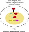
Figure 7. Molecular mechanism for the constitutive nuclear accumulation of MRTF-A/B and their protective roles against endothelial cell injury.
Our present findings are schematically summarized.

Similar articles

Cited by

References

    1. Settleman J. A nuclear MAL-function links Rho to SRF. Mol Cell 11, 1121–1123 (2003). - PubMed
    1. Morita T., Mayanagi T. & Sobue K. Dual roles of myocardin-related transcription factors in epithelial mesenchymal transition via slug induction and actin remodeling. J Cell Biol 179, 1027–1042 (2007). - PMC - PubMed
    1. Iwasaki K., Hayashi K., Fujioka T. & Sobue K. Rho/Rho-associated kinase signal regulates myogenic differentiation via myocardin-related transcription factor-A/Smad-dependent transcription of the Id3 gene. J Biol Chem 283, 21230–21241 (2008). - PMC - PubMed
    1. Mercher T. et al. Involvement of a human gene related to the Drosophila spen gene in the recurrent t(1;22) translocation of acute megakaryocytic leukemia. Proc Natl Acad Sci USA 98, 5776–5779 (2001). - PMC - PubMed
    1. Miralles F., Posern G., Zaromytidou A.I. & Treisman R. Actin dynamics control SRF activity by regulation of its coactivator MAL. Cell 113, 329–342 (2003). - PubMed

Publication types

MeSH terms

LinkOut - more resources