. 2016 Jan;23(1):64-75.
doi: 10.1038/cdd.2015.68. Epub 2015 May 29.

Comprehensive identification of genes driven by ERV9-LTRs reveals TNFRSF10B as a re-activatable mediator of testicular cancer cell death

Affiliations

Comprehensive identification of genes driven by ERV9-LTRs reveals TNFRSF10B as a re-activatable mediator of testicular cancer cell death

U Beyer et al. Cell Death Differ. 2016 Jan.

Abstract

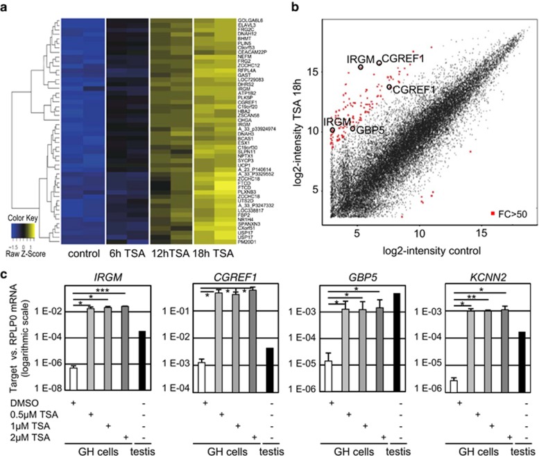
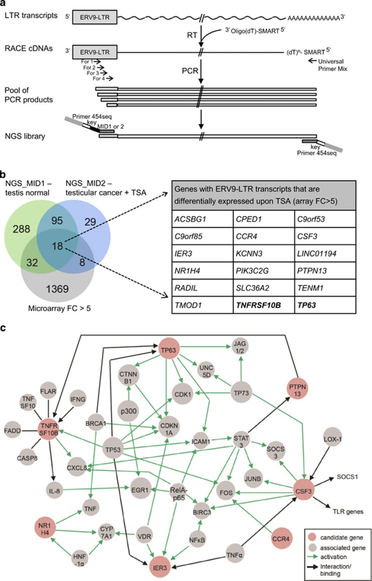
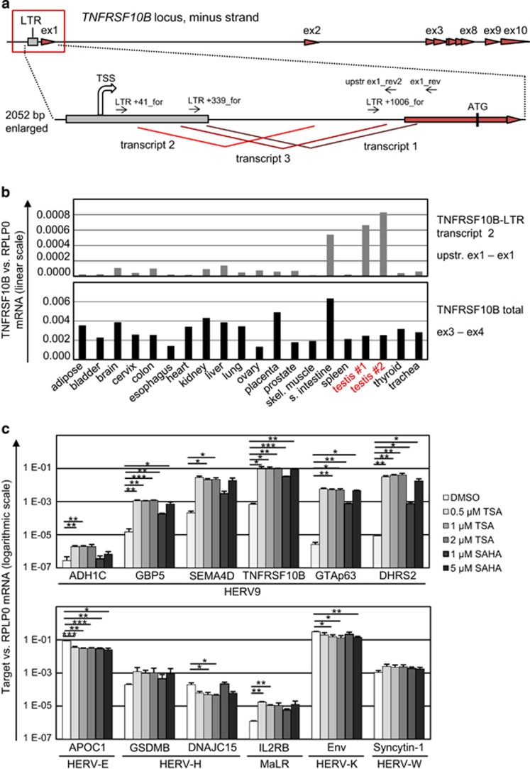
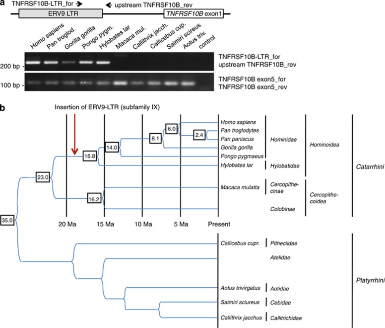
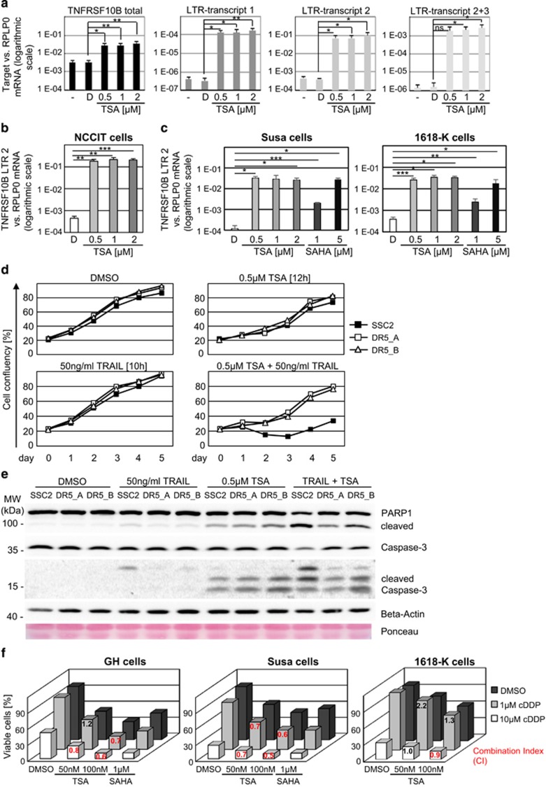
The long terminal repeat (LTR) of human endogenous retrovirus type 9 (ERV9) acts as a germline-specific promoter that induces the expression of a proapoptotic isoform of the tumor suppressor homologue p63, GTAp63, in male germline cells. Testicular cancer cells silence this promoter, but inhibitors of histone deacetylases (HDACs) restore GTAp63 expression and give rise to apoptosis. We show here that numerous additional transcripts throughout the genome are driven by related ERV9-LTRs. 3' Rapid amplification of cDNA ends (3'RACE) was combined with next-generation sequencing to establish a large set of such mRNAs. HDAC inhibitors induce these ERV9-LTR-driven genes but not the LTRs from other ERVs. In particular, a transcript encoding the death receptor DR5 originates from an ERV9-LTR inserted upstream of the protein coding regions of the TNFRSF10B gene, and it shows an expression pattern similar to GTAp63. When treating testicular cancer cells with HDAC inhibitors as well as the death ligand TNF-related apoptosis-inducing ligand (TRAIL), rapid cell death was observed, which depended on TNFRSF10B expression. HDAC inhibitors also cooperate with cisplatin (cDDP) to promote apoptosis in testicular cancer cells. ERV9-LTRs not only drive a large set of human transcripts, but a subset of them acts in a proapoptotic manner. We propose that this avoids the survival of damaged germ cells. HDAC inhibition represents a strategy of restoring the expression of a class of ERV9-LTR-mediated genes in testicular cancer cells, thereby re-enabling tumor suppression.


Figures

Figure 1
Figure 1
Induction of ERV9-LTR-associated genes by HDAC inhibition in testicular cancer cells. (a) Induction of gene expression in testicular cancer cells upon HDAC inhibition. Testicular tumor-derived cells of the line GH were treated with 500 nM TSA for the indicated periods of time. Treatment with the solvent DMSO served as control. mRNA levels were determined by array hybridization. The heatmap shows the top 50 genes that were upregulated upon TSA treatment. Results from two independent experiments are shown. For every displayed gene, the Z-score was calculated: blue, the measured intensity of the sample is lower than the mean intensity of all samples; yellow, the measured intensity of the sample is higher than the mean intensity of all samples. Normalized expression data are shown in Supplementary Table S2. (b) Specific genes are upregulated by more than 50-fold. The scatter plot depicts mean log2 intensities from the oligonucleotides on the array, each corresponding to a specific gene, comparing TSA-induced cells with control cells from the experiment described in (a). Red, differentially expressed transcripts with a fold change (FC) of at least 50; circles, genes that are situated close to an ERV9-LTR in the genome (Supplementary Table S1). Logarithmic FC values of differentially expressed genes upon 18 h of TSA are shown in Supplementary Table S3. (c) Expression of candidate ERV9-LTR-driven genes (of (b) and Supplementary Table S1; array data are shown in Supplementary Tables S2 and S3) is enhanced by TSA treatment. GH cells were treated for 18 h with increasing concentrations of TSA or with the solvent DMSO. Quantification of target gene mRNA levels was performed by real-time RT-PCR, normalized to RPLP0. Mean expression ratios of three independent experiments. P-values were calculated using Student's t-test (ns: P>0.05, *P<0.05, **P<0.01, ***P<0.001). Expression levels in human normal testes were determined for comparison. Note the logarithmic scale
Figure 2
Figure 2
Identification of human transcripts that start at the ERV9-LTR using 3' RACE and NGS experiments. (a) Experimental design to identify human ERV9-LTR-driven transcripts. Human transcripts with LTR sequences at their 5'-end were first reversely transcribed with a modified oligo(dT) primer, then amplified by RACE-PCR using diverse forward primers specific to the transcribed portion of the ERV9-LTR and the reverse Universal Primer Mix (UPM), obtaining a pool of PCR products that were then analyzed by NGS. The primers to produce the NGS library contain Junior454-specific sequences. MID, multiple identifier: MID1, identifier for RACE products from normal human testis; MID2, identifier for RACE products from TSA-treated testicular cancer cells. (b) The Venn diagram shows the overlap of genes identified by RACE and NGS from testis and from TSA-treated testicular cancer cells, as well as TSA-induced genes (fold change, FC>5) determined using array analyses (Figure 1a). The FC of expression (array hybridization) and the numbers of NGS reads for each candidate gene are listed in Table 1 and the NGS raw data is shown in Supplementary Table S5. While ERV9-LTR transcripts from 433 genes were identified in normal testes, only 150 LTR transcripts were detected in TSA-treated testicular cancer cells. Eighteen TSA-inducible genes harbor ERV9-LTR-derived transcripts and were identified by RACE-NGS, both in testes and in testicular tumor cells. While the tumor suppressor TP63 was previously shown to contain an upstream LTR promoter driving GTAp63 expression in testicular germ cells, an ERV9-LTR-driven transcript was also found to arise from the proapoptotic TNFRSF10B gene and this was chosen for subsequent functional analysis. (c) Interaction network of candidate genes involved in cell signaling. A gene ontology search of the 18 candidate genes (of Figure 2b) identified nine of these genes (red circles) having a role in intracellular signaling pathways. The PROTEOME software generated an interaction network based on PubMed-indexed literature, with genes depicted as circles and interaction between different genes as arrows. Green arrows indicate transactivation of the target gene or activation of the target protein, while black arrows mark other kinds of interactions, e.g., protein binding. Gene products within the network function in apoptosis, directly or indirectly, as exemplified by the p53 family of proteins or the tumor necrosis factor (TNF) superfamily
Figure 3
Figure 3
Expression of the death receptor gene TNFRSF10B, encoding Killer/DR5, from an upstream ERV9-LTR. (a) Architecture of the human TNFRSF10B gene, including 10 exons (red arrowheads). The newly identified ERV9-LTR (grey box) is located upstream of the previously described promoter. The region surrounding the LTR and TNFRSF10B exon 1 is shown with a zoom, depicting three different transcripts that are generated from the LTR promoter and alternatively spliced (red lines). The start site of LTR transcription (TSS) is shown by a large arrow, primers for amplification of distinct TNFRSF10B LTR transcripts are indicated by small arrows. Expression of the known and the three newly identified transcripts all result in the synthesis of the same protein, using the start codon located within exon 1. (b) Quantification of TNFRSF10B mRNA expression in a panel of normal human tissues. Real-time RT-PCR was performed using primers that either bind to the region between the LTR and exon 1 (LTR transcript 2; upper panel), or to exons 3 and 4 (total TNFRSF10B mRNA; lower panel). TNFRSF10B mRNA levels were normalized to RPLP0. While similar levels of total TNFRSF10B mRNA were detected in all tissues, the LTR-driven transcript was mainly detected in two different samples of testicular tissue, and in the small intestine. Oligonucleotide sequences are given in Supplementary Table S4. (c) Induction of HERV-regulated transcription upon HDAC inhibition in GH testicular cancer cells is specific for LTRs belonging to the HERV-9 subfamily. Total RNA was isolated from GH cells treated with the solvent DMSO or increasing concentrations of the HDACis TSA or SAHA. Expression of mRNA levels of diverse HERV promoter-driven genes was quantified by real-time RT-PCR, normalized to RPLPO. HDAC inhibition significantly induced the transcription of six HERV-9 LTR-associated genes (upper panel). In contrast, transcription of genes driven by LTRs of the HERV-E, HERV-H or HERV-K subfamilies was rather downregulated upon HDAC inhibition (lower panel). Mean expression ratios of three independent experiments. P-values were calculated using Student's t-test (ns: P>0.05, *P<0.05, **P<0.01, ***P<0.001). Note the logarithmic scale
Figure 4
Figure 4
Insertion of the ERV9-LTR adjacent to TNFRSF10B in hominoid primates. (a) PCR amplification of the region comprising the ERV9-LTR and TNFRSF10B exon 1 sequences from genomic DNA of primates, yielding a specific product in Hominidae and Hylobatidae but not in other primates. Binding of the primers ‘TNFRSF10B LTR_for' and ‘upstream TNFRSF10B_rev' is shown in the upper scheme; sequences are given in Supplementary Table S4. In contrast, a portion of the TNFRSF10B gene was amplified from all primates. (b) Timing of primate evolution and ERV9-LTR insertion adjacent to TNFRSF10B. The pattern and sequence of the TNFRSF10B-associated ERV9-LTR in primate genomes indicates that the insertion of the ERV9-LTR upstream of TNFRSF10B occurred in the ancestor of Hominoidea, during the time prior to Hominidae separation from Hylobatidae, roughly 18 million years ago
Figure 5
Figure 5
TNFRSF10B expression contributes to apoptosis of testicular cancer cells upon HDAC inhibition. (a) HDAC inhibition increases the expression of TNFRSF10B from the LTR promoter, as detected by quantitative real-time RT-PCR. Total RNA was isolated from GH cells treated for 18 h with the indicated concentrations of TSA or DMSO as control. cDNAs were amplified with primers corresponding to total TNFSRF10B or the three distinct LTR transcripts. Mean values, normalized to RPLP0, and standard deviations from three independent experiments are shown. P-values were calculated using Student's t-test (ns: P>0.05, *P<0.05, **P<0.01, ***P<0.001). Notably, the relative level of LTR-derived mRNAs are upregulated by several 100-fold upon TSA treatment (logarithmic scale). (b) Strong induction of the TNFRSF10B transcription from the LTR promoter was also seen upon TSA treatment of NCCIT testicular cancer cells, as determined by quantification of the TNFRSF10B LTR transcript 2 mRNA level. (c) TNFRSF10B expression from the LTR promoter was also increased upon treatment of testicular cancer cells with the HDACi SAHA, a clinically used chemotherapeutic drug. Susa or 1618-K testicular cancer cells were treated with increasing concentrations of TSA or SAHA, or the solvent DMSO. Quantification of TNFRSF10B LTR transcript 2 expression by real-time RT-PCR, normalized to RPLP0. Notably, SAHA treatment had a similar effect on LTR-driven transcription as compared with TSA treatment (logarithmic scale). (d) Depletion of DR5 diminished TSA- and TRAIL-induced apoptosis in testicular cancer cells. After transfection of GH cells with siRNAs targeting DR5 or the control scrambled siRNA SSC2, cells were treated with TSA, or the DR5 ligand TRAIL, or a combination of both. Treatment was performed for 12 h (TSA) or 10 h (TRAIL), washed out at day 1, and cell survival upon treatment was assessed by monitoring of cell confluency consecutively for 4 days. A representative result of three independent experiments is shown; the results of the other two experiments are displayed in Supplementary Figure S5E. Only the combination of TSA and TRAIL led to strongly suppressed cell survival, and this was rescued specifically by the knockdown of DR5. (e) Immunoblot analysis of cells transfected and treated with TRAIL and/or TSA, as described in the legend to Figure 5d. Apoptosis was determined by staining PARP1 and cleaved caspase 3. Beta-actin and Ponceau staining served as controls. In comparison with single treatments, the combined treatment with TRAIL and TSA markedly enhanced PARP1 and caspase 3 cleavage. This effect was rescued in DR5-depleted cells. (f) Synergistic effect of combined treatment of testicular cancer cells with the HDACi TSA and cDDP; the latter drug represents the mainstay of current therapy to eliminate testicular cancer. Testicular cancer cells were treated with the solvent DMSO or increasing concentrations of TSA and/or the indicated concentrations of cDDP. Cell viability was determined 24 h post treatment by luminometric ATP quantification. Mean values of three independent experiments are shown. For each sample treated with the combination of TSA and cDDP, the Non-Constant Combination Index (CI) was calculated. Notably, in all cell lines, treatment with TSA synergistically enhanced cell death induced by cDDP treatment (mean CI values indicated in red). CI<1: synergism; CI=1: additive effect; CI>1: antagonism

Similar articles

Cited by

References

    1. Lander ES, Linton LM, Birren B, Nusbaum C, Zody MC, Baldwin J et al. Initial sequencing and analysis of the human genome. Nature 2001; 409: 860–921. - PubMed
    1. Jern P, Coffin JM. Effects of retroviruses on host genome function. Annu Rev Genet 2008; 42: 709–732. - PubMed
    1. Beyer U, Moll-Rocek J, Moll UM, Dobbelstein M. Endogenous retrovirus drives hitherto unknown proapoptotic p63 isoforms in the male germ line of humans and great apes. Proc Natl Acad Sci USA 2011; 108: 3624–3629. - PMC - PubMed
    1. Liu M, Eiden MV. Role of human endogenous retroviral long terminal repeats (LTRs) in maintaining the integrity of the human germ line. Viruses 2011; 3: 901–905. - PMC - PubMed
    1. Di Cristofano A, Strazzullo M, Longo L, La Mantia G. Characterization and genomic mapping of the ZNF80 locus: expression of this zinc-finger gene is driven by a solitary LTR of ERV9 endogenous retroviral family. Nucleic Acids Res 1995; 23: 2823–2830. - PMC - PubMed

Publication types

MeSH terms