. 2015 May 29;10(5):e0128177.
doi: 10.1371/journal.pone.0128177. eCollection 2015.

Exon organization and novel alternative splicing of Ank3 in mouse heart

Affiliations

Exon organization and novel alternative splicing of Ank3 in mouse heart

Gokay Yamankurt et al. PLoS One. .

Abstract

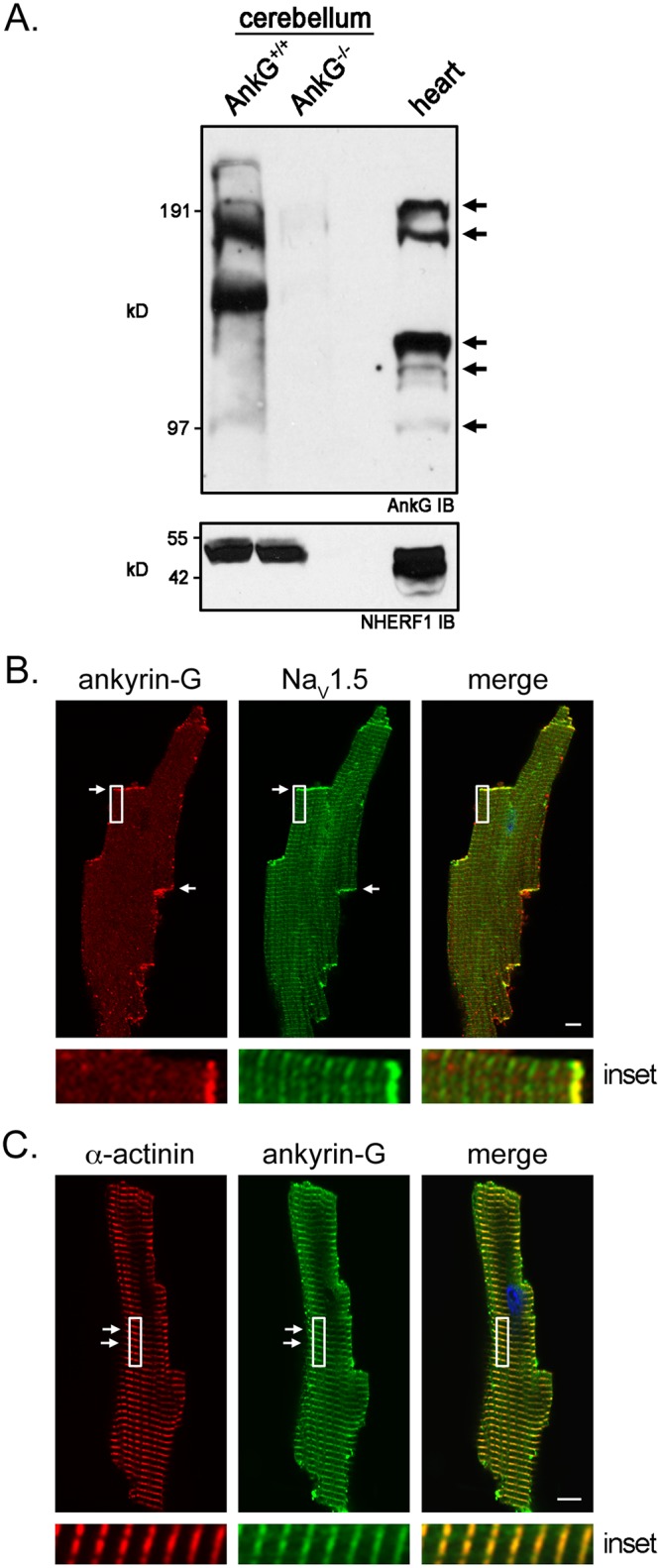
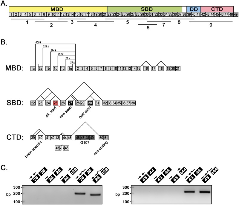
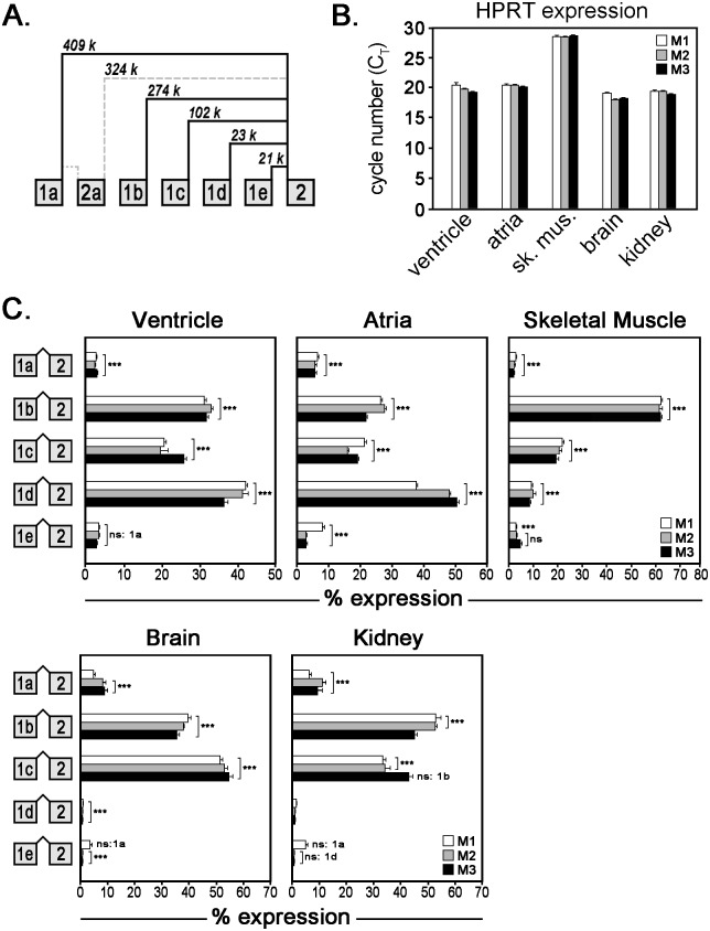
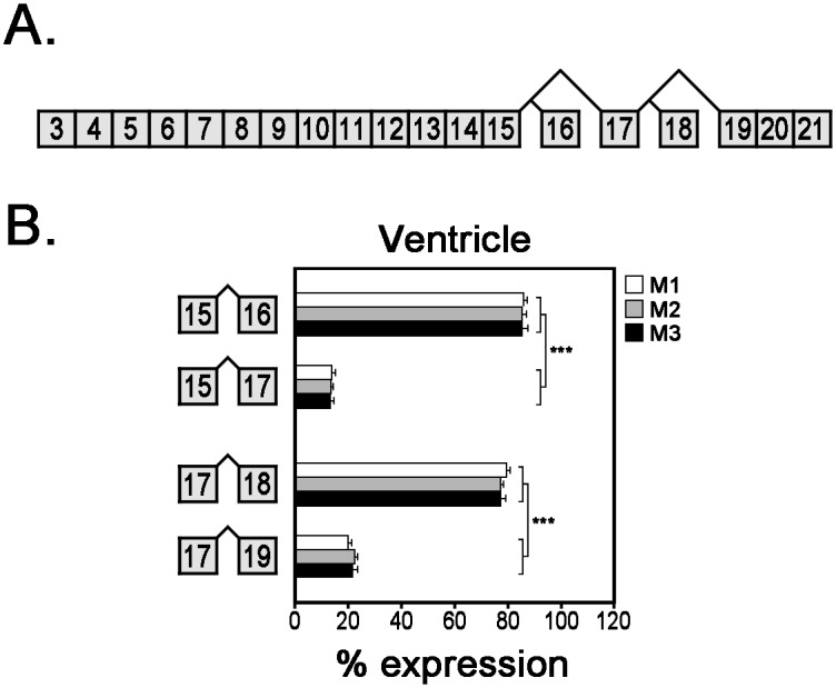
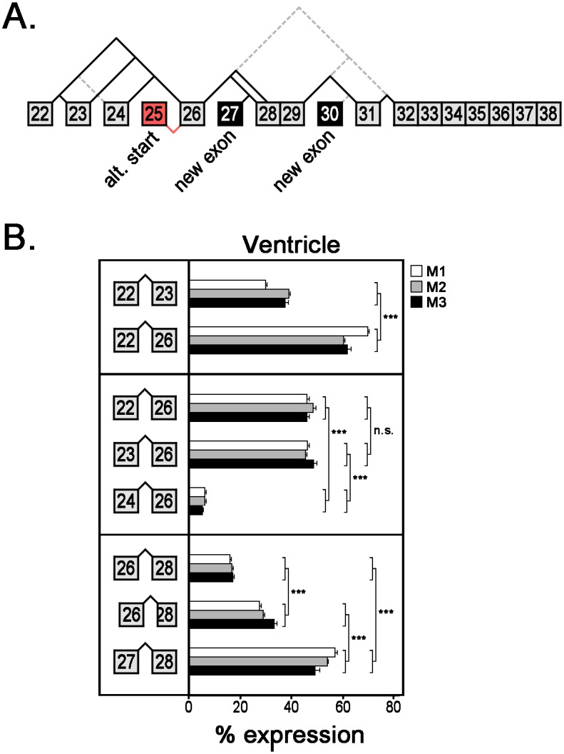
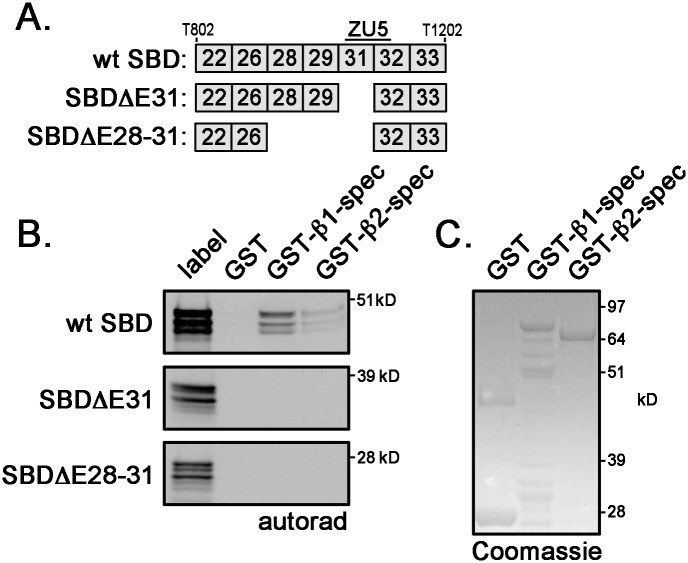
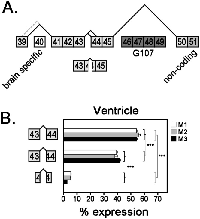
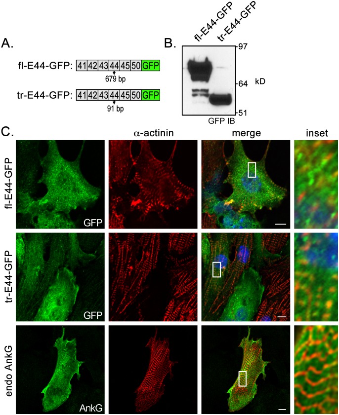
Ankyrin-G is an adaptor protein that links membrane proteins to the underlying cytoskeletal network. Alternative splicing of the Ank3 gene gives rise to multiple ankyrin-G isoforms in numerous tissues. To date, only one ankyrin-G isoform has been characterized in heart and transcriptional regulation of the Ank3 gene is completely unknown. In this study, we describe the first comprehensive analysis of Ank3 expression in heart. Using a PCR-based screen of cardiac mRNA transcripts, we identify two new exons and 28 alternative splice variants of the Ank3 gene. We measure the relative expression of each splice variant using quantitative real-time PCR and exon-exon boundary spanning primers that specifically amplify individual Ank3 variants. Six variants are rarely expressed (<1%), while the remaining variants display similar expression patterns in three hearts. Of the five first exons in the Ank3 gene, exon 1d is only expressed in heart and skeletal muscle as it was not detected in brain, kidney, cerebellum, and lung. Immunoblot analysis reveals multiple ankyrin-G isoforms in heart, and two ankyrin-G subpopulations are detected in adult cardiomyocytes by immunofluorescence. One population co-localizes with the voltage-gated sodium channel NaV1.5 at the intercalated disc, while the other population expresses at the Z-line. Two of the rare splice variants excise a portion of the ZU5 motif, which encodes the minimal spectrin-binding domain, and these variants lack β-spectrin binding. Together, these data demonstrate that Ank3 is subject to complex splicing regulation resulting in a diverse population of ankyrin-G isoforms in heart.


Conflict of interest statement

Competing Interests: The authors have declared that no competing interests exist.

Figures

Fig 1
Fig 1. Immunoblot and immunofluorescent detection of ankyrin-G isoforms in heart.
(A) Ankyrin-G immunoblot demonstrates expression of different isoforms in heart (arrows). Loss of ankyrin-G immunoreactive polypeptides in ankyrin-G null cerebellums demonstrates antibody specificity. NHERF1 immunoblot demonstrates similar loading of protein lysates. (B) Immunofluorescent co-localization of ankyrin-G (monoclonal antibody) and the voltage-gated sodium channel NaV1.5 at intercalated discs of an individually isolated adult cardiomyocyte (white arrows and highlighted in the inset). (C) Immunofluorescent co-localization of ankyrin-G (polyclonal antibody) and α-actinin at Z-lines of individually isolated adult cardiomyocytes (white arrows and highlighted in the inset). Scale bar represents 10 microns.
Fig 2
Fig 2. Exon organization and alternative splicing of Ank3 gene.
(A) Diagram of ankyrin-G exon organization and nine overlapping PCR primer sets used to amplify cardiac-specific Ank3 alternative transcripts. Ankyrin functional domains are indicated. MBD: membrane-binding domain, SBD: spectrin-binding domain, DD: death domain, CTD: C-terminal domain. The large (7694 bp) brain-specific exon corresponds to exon 38. (B) Diagram of alternative Ank3 splice variants encoding the MBD, SBD, and CTD. In the MBD, 5 alternative first exons (1a-1e) were detected. Lengths of intervening intronic sequences are labeled in kilobases (k). In the SBD, two novel exons (27 and 30) were identified and exon numbering has been adjusted accordingly (exon 40 is now the brain-specific exon). Exon 25 (in red) encodes the alternative start site for truncated ankyrin-G isoforms that lack MBDs (e.g. AnkG-107). In the CTD, alternative splicing of exons 46–49 (highlighted in gray) was not addressed in this study. (C) Full-length exon-exon boundary spanning primers are necessary to amplify PCR products: Ank3 splice junctions of exons 26/28 and 26/t28 (truncated) (first panel) and exons 43/44 and 43/t44 (second panel).
Fig 3
Fig 3. Relative mRNA expression of alternative Ank3 first exons.
(A) Diagram of alternative splicing of 5 first exons of the Ank3 gene. Gray dashed lines represent rare alternative spliced junctions (<1%). (B) qt-PCR analysis of hypoxanthine-guanine phosphoribosyltransferase (HPRT) expression (measured as cycle threshold number) in ventricle, atria, skeletal muscle, brain, and kidney from three mice (labeled M1–M3). (C) Relative mRNA expression of alternative first exons was measured in heart (ventricle and atria), skeletal muscle, brain, and kidney by qt-PCR analysis. Bar graphs represent technical replicates of qt-PCR samples and error bars represent standard deviations. Statistical analysis was performed with one-way ANOVA with Tukey’s multiple comparison test (*** p-value ≤0.001, ns: not significant) to assess the significance of expression differences between different splice variants (i.e. 1a vs. 1b).
Fig 4
Fig 4. Relative mRNA expression of alternative Ank3 transcripts encoding ankyrin-G MBD.
(A) Diagram of Ank3 alternative splicing in ankyrin-G MBD. (B) Expression of Ank3 mRNA transcripts ± exon 16 or ± exon 18 was measured in three mouse hearts using qt-PCR analysis with exon-exon boundary spanning primers. Bar graphs represent technical replicates of qt-PCR samples and error bars represent standard deviations. Statistical analysis was performed with unpaired Student’s T-test (*** p-value ≤0.001).
Fig 5
Fig 5. Relative mRNA expression of alternative Ank3 transcripts encoding ankyrin-G SBD.
(A) Diagram of Ank3 alternative splicing in ankyrin-G SBD. Newly identified exons are labeled in black and the alternative first exon (E25) is labeled in red. Gray dashed lines represent rare alternative spliced junctions (<1%). (B) Expression of various Ank3 mRNA transcripts was measured in three mouse hearts using qt-PCR analysis with exon-exon boundary spanning primers. Bar graphs represent technical replicates of qt-PCR samples and error bars represent standard deviations. Statistical analysis was performed using one-way ANOVA with Tukey’s multiple comparison test or unpaired Student’s T-test (*** p-value ≤0.001, ns: not significant).
Fig 6
Fig 6. Alternative ankyrin-G SBD isoforms do not bind β-spectrin.
(A) Diagram of Ank3 exon organizations for two rare ankyrin-G SBD variants (ΔE31 and ΔE28–31). Both variants lack a portion of the ZU5 motif. (B) β1-spectrin and β2-spectrin (repeats 13 to 17) binding to ankyrin-G SBDs (wildtype, Δ31, Δ28–31) was measured by in vitro binding assays. Label is the unbound in vitro translated SBD products. (C) Coomassie Blue Stain demonstrates equal loading of GST-fusion proteins β1- and β2-spectrins.
Fig 7
Fig 7. Relative mRNA expression of alternative Ank3 transcripts encoding ankyrin-G CTD.
(A) Diagram of Ank3 alternative splicing in ankyrin-G CTD. Exon 40 is the large (7694 bp) brain-specific exon, while exon 51 contains non-coding sequence. Exons 46–49 are expressed in truncated ankyrin-G isoforms that lack membrane-binding domains. Gray dashed lines represent rare alternative spliced junctions (<1%). (B) Expression of various Ank3 mRNA transcripts with different iterations of exon 44 (full-length, 5’-truncated, medially truncated) was measured in three mouse hearts using qt-PCR analysis with exon-exon boundary spanning primers. Bar graphs represent technical replicates of qt-PCR samples and error bars represent standard deviations. Statistical analysis was performed with one-way ANOVA with Tukey’s multiple comparison test (*** p-value ≤0.001).
Fig 8
Fig 8. Subcellular localization of ankyrin-G CTD isoforms in neonatal cardiomyocytes.
(A) Diagram of lenti-viral GFP-tagged CTD variants with full-length or truncated exon 44. (B) GFP immunoblot analysis demonstrating expression of lenti-viral constructs. (C) Immunofluorescent co-localization of GFP-tagged CTD variants (fl-E44-GFP and tr-E44-GFP) with Z-line resident protein α-actinin in neonatal cardiomyocytes. Bottom panels represent localization of endogenous ankyrin-G in reference to α-actinin. Scale bar represents 10 microns.

Similar articles

Cited by

References

    1. Devarajan P, Stabach PR, Mann AS, Ardito T, Kashgarian M, Morrow JS. Identification of a small cytoplasmic ankyrin (AnkG119) in the kidney and muscle that binds beta I sigma spectrin and associates with the Golgi apparatus. The Journal of cell biology. 1996;133(4):819–30. - PMC - PubMed
    1. Gagelin C, Constantin B, Deprette C, Ludosky MA, Recouvreur M, Cartaud J, et al. Identification of Ank(G107), a muscle-specific ankyrin-G isoform. The Journal of biological chemistry. 2002;277(15):12978–87. 10.1074/jbc.M111299200 . - DOI - PubMed
    1. Hopitzan AA, Baines AJ, Ludosky MA, Recouvreur M, Kordeli E. Ankyrin-G in skeletal muscle: tissue-specific alternative splicing contributes to the complexity of the sarcolemmal cytoskeleton. Experimental cell research. 2005;309(1):86–98. 10.1016/j.yexcr.2005.04.013 . - DOI - PubMed
    1. Kordeli E, Ludosky MA, Deprette C, Frappier T, Cartaud J. AnkyrinG is associated with the postsynaptic membrane and the sarcoplasmic reticulum in the skeletal muscle fiber. Journal of cell science. 1998;111 (Pt 15):2197–207. . - PubMed
    1. Thevananther S, Kolli AH, Devarajan P. Identification of a novel ankyrin isoform (AnkG190) in kidney and lung that associates with the plasma membrane and binds alpha-Na, K-ATPase. The Journal of biological chemistry. 1998;273(37):23952–8. Epub 1998/09/03. . - PubMed

Publication types

LinkOut - more resources