. 2015 May 29;10(5):e0128489.
doi: 10.1371/journal.pone.0128489. eCollection 2015.

Cell kinetic studies fail to identify sequentially proliferating progenitors as the major source of epithelial renewal in the adult murine prostate

Affiliations

Cell kinetic studies fail to identify sequentially proliferating progenitors as the major source of epithelial renewal in the adult murine prostate

Jean-Christophe Pignon et al. PLoS One. .

Abstract

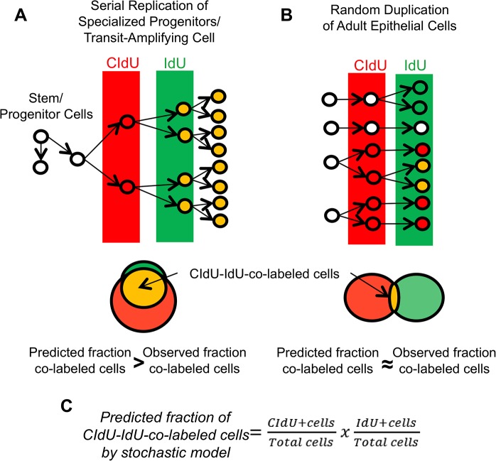
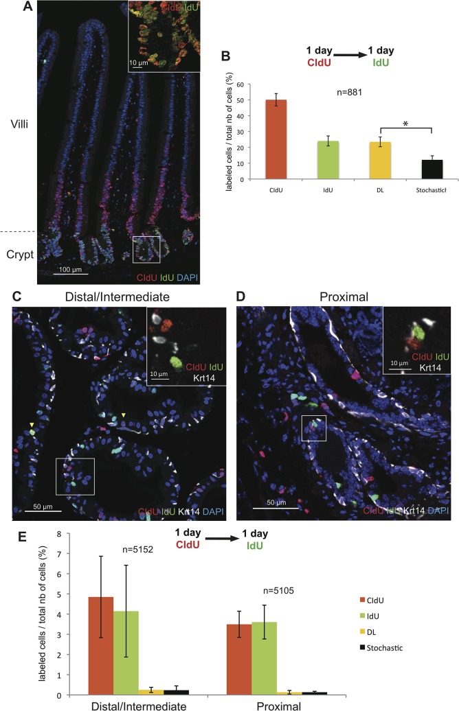
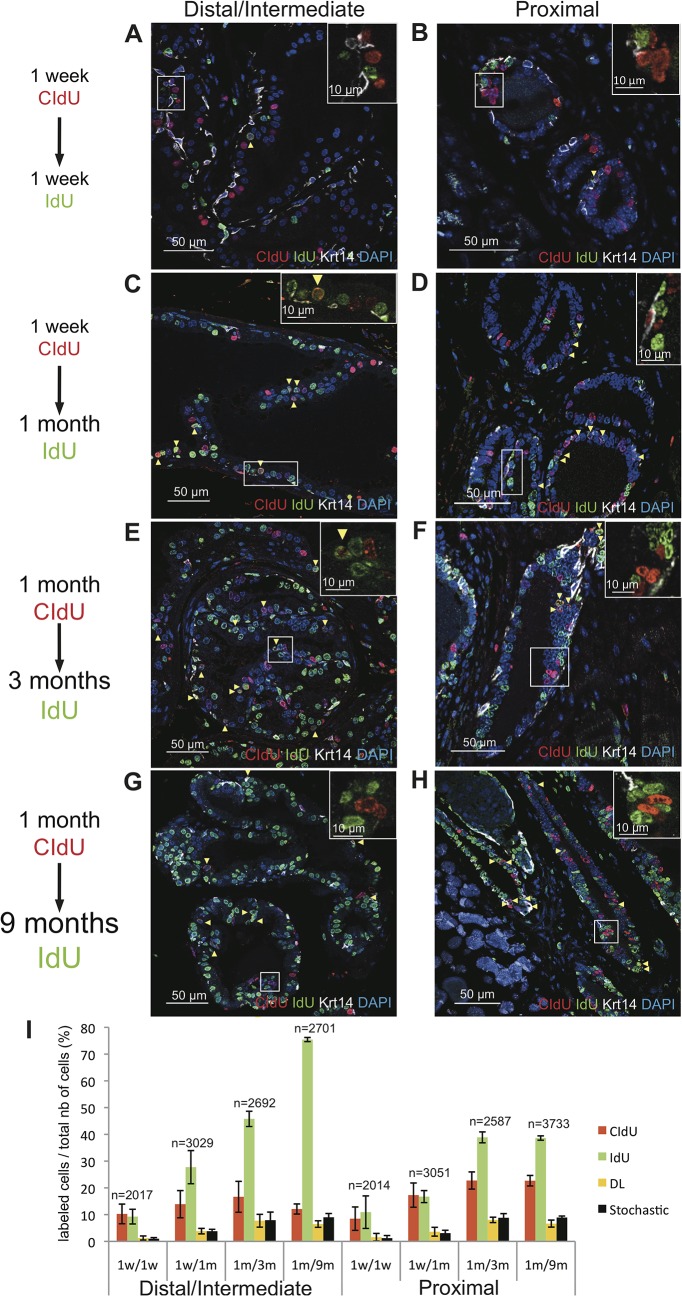
There is evidence that stem cells and their progeny play a role in the development of the prostate. Although stem cells are also considered to give rise to differentiated progeny in the adult prostate epithelium ex vivo, the cohort of adult prostate stem cells in vivo as well as the mechanisms by which the adult prostate epithelium is maintained and regenerated remain highly controversial. We have attempted to resolve this conundrum by performing in vivo tracing of serially replicating cells after the sequential administration of two thymidine analogues to mice. Our results show that, during normal prostate homeostasis, sequentially proliferating cells are detected at a rate that is consistent with a stochastic process. These findings indicate that in vivo, under steady-state conditions, most adult prostate epithelial cells do not represent the progeny of a small number of specialized progenitors that generate sequentially replicating transit-amplifying (TA) cells but are formed by stochastic cell division. Similarly, no rapidly cycling TA cells were detected during regeneration following one cycle of androgen-mediated involution/regeneration of the prostate epithelium. These findings greatly enhance our understanding of the mechanisms regulating prostate epithelial cell renewal and may have significant implications in defining the cell of origin of proliferative prostatic diseases.


Conflict of interest statement

Competing Interests: The authors have declared that no competing interests exist.

Figures

Fig 1
Fig 1. Experimental design.
Consecutive administration of two different thymidine analogs, namely CIdU (in red) and IdU (in green), to mice allows the detection of serially replicating cells (CIdU-IdU-co-labeled cells, in orange). (A): According to the experimental methodology developed by Teta et al. [3], if the prostate epithelium is maintained by transit-amplifying (TA) cells that undergo consecutive cycles of cell division, a significant number of proliferating cells would be co-labeled by both thymidine analogs. In that case, the observed fraction of co-labeled cells should be greater than predicted fraction of co-labeled cells by the stochastic model. (B): In contrast, if the epithelium is maintained by cell division that occurs stochastically, the majority of replicating cells should be labeled with either one or the other thymidine analog. In this situation, the observed fraction of co-labeled cells should similar to the fraction of co-labeled cells predicted by the stochastic model. (C): The fraction of co-labeled cells predicted by the stochastic model is calculated by multiplying the fraction of CIdU-labeled cells by the fraction of IdU-labeled cells. This figure has been adapted from Humphreys et al. [8].
Fig 2
Fig 2. Renewal of the adult prostate epithelium does not depend on rapidly serially proliferating progenitor/TA cells.
Detection of rapidly proliferating progenitors was performed on 7-week-old mice sequentially treated with CIdU and IdU for 1 day each. Mice were sacrificed immediately after the end of IdU administration. (A): Representative images of double immunofluorescence staining for CIdU and IdU in a section of small intestine demonstrating the presence of double-labeled progenitor cells in the intestinal crypt (inset). (B): Graphic representation of the percentages of intestinal epithelial cells labeled with CIdU, IdU, or CIdU/IdU. The predicted stochastic fraction is also shown. Data represent the means ± SD for three mice per group. (C, D): Representative images of triple immunofluorescence staining for CIdU, IdU and Krt14 performed on sections of the distal/intermediate region of ducts (C) and the proximal region of ducts (D) of the dorsal prostate showing that the majority of cells are single labeled (inset). Yellow arrowheads indicate CIdU-IdU-co-labeled cells. (E): Graphic representation of the percentages of epithelial prostate cells (both basal and luminal) labeled with CIdU, IdU, or CIdU/IdU (DL). The predicted stochastic fraction is also shown. Data represent the means ± SD for three mice per group. n indicates the average number of nuclei counted per mouse.* indicates p<0.05.
Fig 3
Fig 3. Renewal of the adult prostate epithelium does not depend on slowly serially proliferating progenitor/TA cells.
Representative images of triple immunofluorescence staining for CIdU, IdU and Krt14 on sections of the distal/intermediate region (A, C, E, G) and the proximal region of ducts (B, D, F, H) of the dorsal prostate of 7-week-old mice treated for either 1 week with CIdU followed by 1 week of IdU (A, B), or 1 week with CIdU followed by 1 month of IdU (C, D), or for 1 month of CIdU followed by 3 months of IdU (E, F), or for 1 month of CIdU followed by 9 months of IdU (G, H). In all experiments, mice were sacrificed immediately after the end of IdU administration. Yellow arrowheads indicate double-labeled cells while insets show that the majority of the cells are single labeled. (I): Graphic representation of the percentages of epithelial prostate cells (both basal and luminal) labeled with CIdU, IdU, or CIdU/IdU (DL) for the indicated treatment groups. The predicted stochastic fraction is also shown. Data represent the means ± SD for three mice per group. n indicates the average number of nuclei counted per mouse.
Fig 4
Fig 4. Androgen mediated regeneration of the prostate epithelium after castration does not depend on rapidly serially proliferating progenitor/TA cells.
Representative images of triple immunofluorescence staining for CIdU, IdU and Krt14 on sections of the distal/intermediate (A, C) and the proximal (B, D) regions of ducts from the dorsal prostate of castrated mice sequentially treated with CIdU and IdU (1 day each) at day 2 (A, B) or day 3 (C, D) after androgen supplementation. In all experiments, mice were sacrificed immediately after the end of IdU administration. Yellow arrowheads indicate double-labeled cells while insets show that the majority of the cells are single labeled. (E): Graphic representation of the percentages of epithelial prostate cells (both basal and luminal) labeled with CIdU, IdU, or CIdU/IdU (DL) for the indicated treatment groups. 2dR and 3dR indicate mice that were treated with the thymidine analogs at day 2 or day 3 after androgen supplementation, respectively. The predicted stochastic fraction is also shown. Data represent the means ± SD for three mice per group. n indicates the average number of nuclei counted per mouse. * indicates p<0.05.

References

    1. Dor Y, Brown J, Martinez OI, Melton DA. Adult pancreatic beta-cells are formed by self-duplication rather than stem-cell differentiation. Nature. 2004;429:41–46. - PubMed
    1. Senyo SE, Steinhauser ML, Pizzimenti CL, Yang VK, Cai L, Wang M, et al. Mammalian heart renewal by pre-existing cardiomyocytes. Nature. 2013;493:433–436. 10.1038/nature11682 - DOI - PMC - PubMed
    1. Teta M, Rankin MM, Long SY, Stein GM, Kushner JA. Growth and regeneration of adult beta cells does not involve specialized progenitors. Dev Cell. 2007;12:817–826. - PubMed
    1. Ousset M, Van Keymeulen A, Bouvencourt G, Sharma N, Achouri Y, Simons BD, et al. Multipotent and unipotent progenitors contribute to prostate postnatal development. Nat Cell Biol. 2012;14:1131–1138. 10.1038/ncb2600 - DOI - PubMed
    1. Pignon JC, Grisanzio C, Geng Y, Song J, Shivdasani RA, Signoretti S. p63-expressing cells are the stem cells of developing prostate, bladder, and colorectal epithelia. Proc Natl Acad Sci U S A. 2013;110:8105–8110. 10.1073/pnas.1221216110 - DOI - PMC - PubMed

Publication types