

Immunohistochemical Mapping of Sensory Nerve Endings in the Human Triangular Fibrocartilage Complex
- PMID: 26024577
- PMCID: PMC4562925
- DOI: 10.1007/s11999-015-4357-z
Immunohistochemical Mapping of Sensory Nerve Endings in the Human Triangular Fibrocartilage Complex
Abstract
Background: The triangular fibrocartilage complex is the main stabilizer of the distal radioulnar joint. While static joint stability is constituted by osseous and ligamentous integrity, the dynamic aspects of joint stability chiefly concern proprioceptive control of the compressive and directional muscular forces acting on the joint. Therefore, an investigation of the pattern and types of sensory nerve endings gives more insight in dynamic distal radioulnar joint stability.
Questions/purposes: We aimed to (1) analyze the general distribution of sensory nerve endings and blood vessels; (2) examine interstructural distribution of sensory nerve endings and blood vessels; (3) compare the number and types of mechanoreceptors in each part; and (4) analyze intrastructural distribution of nerve endings at different tissue depth.
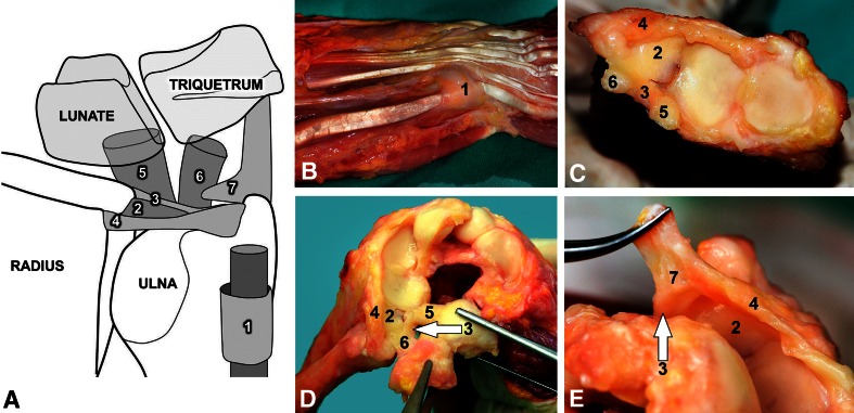
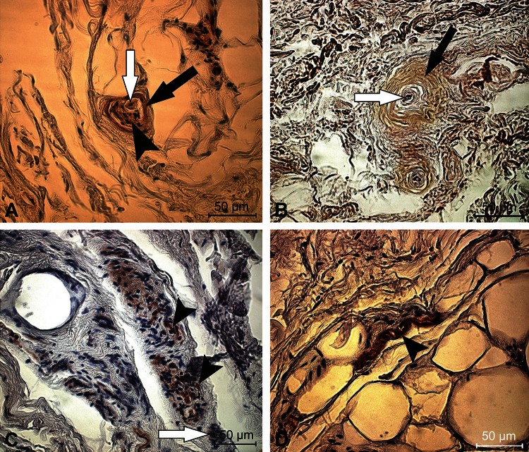
Methods: The subsheath of the extensor carpi ulnaris tendon sheath, the ulnocarpal meniscoid, the articular disc, the dorsal and volar radioulnar ligaments, and the ulnolunate and ulnotriquetral ligaments were dissected from 11 human cadaver wrists. Sensory nerve endings were counted in five levels per specimen as total cell amount/cm(2) after staining with low-affinity neurotrophin receptor p75, protein gene product 9.5, and S-100 protein and thereafter classified according to Freeman and Wyke.
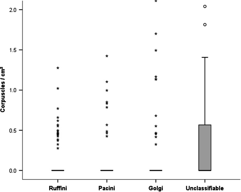
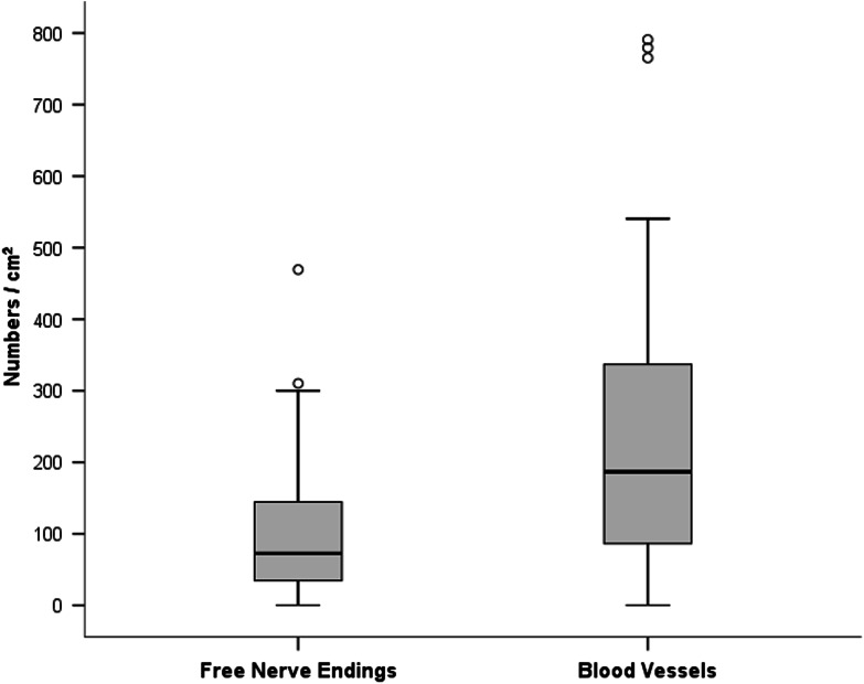
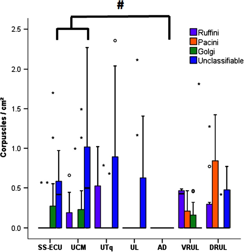
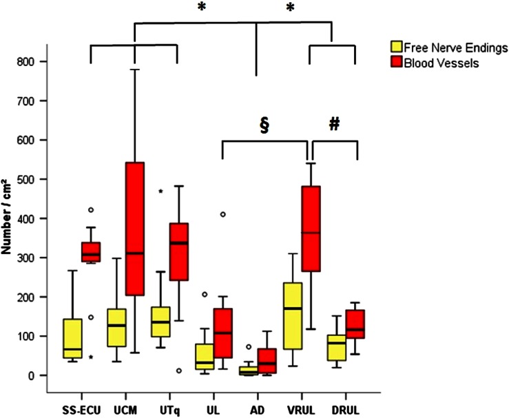
Results: All types of sensory corpuscles were found in the various structures of the triangular fibrocartilage complex with the exception of the ulnolunate ligament, which contained only Golgi-like endings, free nerve endings, and unclassifiable corpuscles. The articular disc had only free nerve endings. Furthermore, free nerve endings were the predominant sensory nerve ending (median, 72.6/cm(2); range, 0-469.4/cm(2)) and more prevalent than all other types of mechanoreceptors: Ruffini (median, 0; range, 0-5.6/cm(2); difference of medians, 72.6; p < 0.001), Pacini (median, 0; range, 0-3.8/cm(2); difference of medians, 72.6; p < 0.001), Golgi-like (median, 0; range, 0-2.1/cm(2); difference of medians, 72.6; p < 0.001), and unclassifiable corpuscles (median, 0; range, 0-2.5/cm(2); difference of medians, 72.6; p < 0.001). The articular disc contained fewer free nerve endings (median, 1.8; range, 0-17.8/cm(2)) and fewer blood vessels (median, 29.8; range, 0-112.2/cm(2); difference of medians: 255.9) than all other structures of the triangular fibrocartilage complex (p ≤ 0.001, respectively) except the ulnolunate ligament. More blood vessels were seen in the volar radioulnar ligament (median, 363.62; range, 117.8-871.8/cm(2)) compared with the ulnolunate ligament (median, 107.7; range, 15.9-410.3/cm(2); difference of medians: 255.91; p = 0.002) and the dorsal radioulnar ligament (median, 116.2; range, 53.9-185.1/cm(2); difference of medians: 247.47; p = 0.001). Free nerve endings were obtained in each structure more often than all other types of sensory nerve endings (p < 0.001, respectively). The intrastructural analysis revealed no differences in mechanoreceptor distribution in all investigated specimens with the numbers available, showing a homogenous distribution of proprioceptive qualities in all seven parts of the triangular fibrocartilage complex.
Conclusions: Nociception has a primary proprioceptive role in the neuromuscular stability of the distal radioulnar joint. The articular disc and ulnolunate ligament rarely are innervated, which implies mainly mechanical functions, whereas all other structures have pronounced proprioceptive qualities, prerequisite for dynamic joint stability.
Clinical relevance: Lesions of the volar and dorsal radioulnar ligaments have immense consequences not only for mechanical but also for dynamic stability of the distal radioulnar joint, and surgical reconstruction in instances of radioulnar ligament injury is important.
Figures
References
-
- Cavalcante ML, Rodrigues CJ, Mattar R Jr. Mechanoreceptors and nerve endings of the triangular fibrocartilage in the human wrist. J Hand Surg Am. 2004;29:432–435; discussion 436–438. - PubMed
-
- Frank CB. Ligament structure, physiology and function. J Musculoskelet Neuronal Interact. 2004;4:199–201. - PubMed
-
- Garcia-Elias M. Soft-tissue anatomy and relationships about the distal ulna. Hand Clin. 1998;14:165–176. - PubMed
Publication types
MeSH terms
LinkOut - more resources
Full Text Sources
Other Literature Sources
Research Materials
