Review
. 2015 Aug 1;309(3):H381-92.
doi: 10.1152/ajpheart.00077.2015. Epub 2015 May 29.

Neural control of circulation and exercise: a translational approach disclosing interactions between central command, arterial baroreflex, and muscle metaboreflex

Affiliations
Review

Neural control of circulation and exercise: a translational approach disclosing interactions between central command, arterial baroreflex, and muscle metaboreflex

Lisete C Michelini et al. Am J Physiol Heart Circ Physiol. .

Abstract

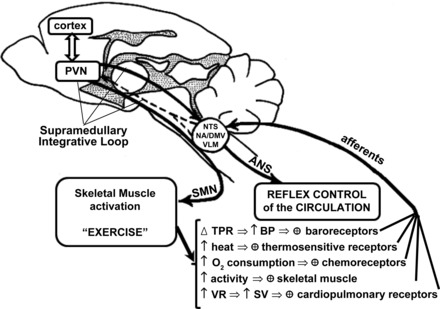
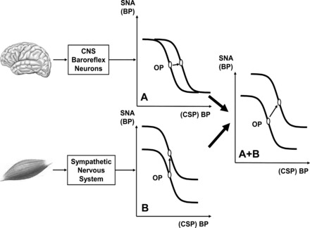
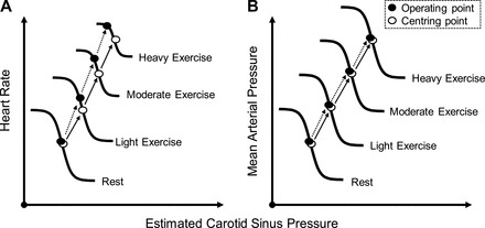
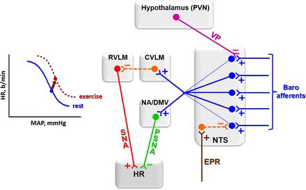
The last 100 years witnessed a rapid and progressive development of the body of knowledge concerning the neural control of the cardiovascular system in health and disease. The understanding of the complexity and the relevance of the neuroregulatory system continues to evolve and as a result raises new questions. The purpose of this review is to articulate results from studies involving experimental models in animals as well as in humans concerning the interaction between the neural mechanisms mediating the hemodynamic responses during exercise. The review describes the arterial baroreflex, the pivotal mechanism controlling mean arterial blood pressure and its fluctuations along with the two main activation mechanisms to exercise: central command (parallel activation of central somatomotor and autonomic descending pathways) and the muscle metaboreflex, the metabolic component of exercise pressor reflex (feedback from ergoreceptors within contracting skeletal muscles). In addition, the role of the cardiopulmonary baroreceptors in modulating the resetting of arterial baroreflex is identified, and the mechanisms in the central nervous system involved with the resetting of baroreflex function during dynamic exercise are also described. Approaching a very relevant clinical condition, the review also presents the concept that the impaired arterial baroreflex function is an integral component of the metaboreflex-mediated exaggerated sympathetic tone in subjects with heart failure. This increased sympathetic activity has a major role in causing the depressed ventricular function observed during submaximal dynamic exercise in these patients. The potential contribution of a metaboreflex arising from respiratory muscles is also considered.

Keywords: autonomic; baroreflex; central command; exercise; metaboreflex.


Figures

Fig. 1.
Fig. 1.
Schematic representation of somatomotor and autonomic outflows controlling muscle activity and the reflex control of the circulation. Different receptors detect changes in muscle contraction/perfusion, cardiac filling, blood pressure (BP), blood gases, and temperature during muscular activity and send this information to brain stem and supramedullary areas involved in reflex control of the circulation. ANS, autonomic nervous system; DMV, dorsomotor nucleus of the vagus; NA, nucleus ambiguous; NTS, nucleus of the solitary tract; PVN, paraventricular nucleus of the hypothalamus; SMN, somatomotor neurons; SV, stroke volume; TPR, total peripheral resistance; VLM, ventrolateral medulla; VR, venous return. Modified from Fig. 3 in Michelini and Morris (68).
Fig. 2.
Fig. 2.
Hypothetical stimulus-response curves for arterial baroreflex, expressed as relationship between sympathetic nervous activity (SNA) and systemic BP or often in experiments as relationship between BP and isolated carotid sinus pressure (CSP). Operating point (OP) is BP “sought” by arterial baroreflex (often point of maximal gain). A: resetting of baroreflex by central command, a stimulus acting on neuron pool receiving baroreceptor afferent. OP is shifted laterally to a higher BP. CNS, central nervous system. B: vertical shift of baroreflex function curve by muscle chemoreflex, a stimulus that increases SNA and raises BP without changing OP; i.e., influence is confined to efferent arm of baroreflex. A + B: hypothetical combined effects of both stimuli on curve during exercise (upward and rightward resetting). Modified from Fig. 4 in Rowell and O'Leary (95).
Fig. 3.
Fig. 3.
Representative illustration of the intensity-dependent resetting of the carotid baroreflex during dynamic exercise of 7 subjects. The centering point (CP) is the point at which there is an equal depressor and pressor response to a given change in BP; the OP is the prestimulus BP. Both the carotid baroreflex-heart rate (HR; A) and mean arterial pressure (MAP; B) stimulus-response curves progressively reset during exercise in an intensity-dependent manner without significant changes in maximal gain (sensitivity). A consistent observation for the baroreflex control of HR is the relocation of the OP away from the CP and closer to the threshold of the stimulus-response curve along with a reduction in the response range as exercise intensity increases (A). The relocation of the OP for HR control positions the baroreflex in a more optimal position to counter hypertensive stimuli with increasing exercise intensity. In contrast, for the carotid baroreflex-MAP, stimulus-response curve, the OP does not relocate away from the CP and the response range remains the same as at rest with an upward and rightward shift in parallel with an increase in the intensity of exercise (B). Previously published in Fadel and Raven (29).
Fig. 4.
Fig. 4.
Proposed interaction between arterial baroreceptor afferents, exercise pressor reflex (EPR), and PVN vasopressinergic (VP) pathways that project to second-order neurons within the NTS. During exercise, both EPR (through a GABAergic projection) and VP (through an axo-axonal inhibitory synapse) partially inhibit afferent signaling by baroreceptors, thus limiting the degree of baroinhibition of the heart during pressure increases. These exercise-induced effects instantaneously reset baroreflex function (left). Blue, excitatory glutamatergic pathways; orange, GABAergic inhibitory pathways; pink, descending vasopressinergic projections; green, parasympathetic nervous activity (PSNA); red, SNA to the heart. CVLM, caudal ventrolateral medulla; RVLM, rostral ventrolateral medulla. b/min, Beats/min.
Fig. 5.
Fig. 5.
Graphs depicting the effect of exercise on the activation of descending PVN-NTS vasopressinergic projections [top left: modified from Fig. 4 in Michelini (66)] and the functional responses induced by NTS administration of vasopressin on baroreceptor reflex control of HR [top right: redrawn from data published in Michelini and Bonagamba (67)] and by NTS VP antagonist blockade on HR response during graded exercise on treadmill [bottom: reproduced with permission from Michelini and Stern (69)]. S and T, sedentary and trained groups, respectively; Veh, vehicle; VP, vasopressin; VPant, VP antagonist. The parameters for sigmoidal fitting are as follows: VehNTS: inferior plateau = 242 ± 10 beats/min; range = 232 ± 17 beats/min; blood pressure at half of the HR range = 101 ± 3 mmHg; gain = −3.55 ± 0.45 beats·min−1·mmHg−1; VPNTS: inferior plateau = 293 ± 9 beats/min*; range = 192 ± 15 beats/min*; blood pressure at half of the HR range = 116 ± 2 mmHg*; gain = −4.05 ± 0.48 beats·min−1·mmHg−1. *P < 0.05 vs. Veh treatment; +P < 0.05 vs. rest;·P < 0.05 vs. S group.
Fig. 6.
Fig. 6.
Relationship between hindlimb blood flow and HR during mild and moderate exercise (Ex) before (solid lines) and after (dashed lines) induction of heart failure. Hindlimb blood flow was reduced via transient vascular occlusion of the terminal aorta. During mild exercise, a clear threshold exists for metaboreflex activation both before and after induction of heart failure as hindlimb blood flow must be reduced below threshold from the free flow (no occlusion) levels before tachycardia occurs. However, during moderate exercise in heart failure, the free flow level of hindlimb blood flow (indicated by vertical dashed lines) is now below the threshold seen in control experiments, indicating that the reflex is now tonically active [modified from Hammond et al. (35)].

Similar articles

Cited by

References

    1. Amann M, Blain GM, Proctor LT, Sebranek JJ, Pegelow DF, Dempsey JA. Group III and IV muscle afferents contribute to ventilatory and cardiovascular response to rhythmic exercise in humans. J Appl Physiol (1985) 109: 966–976, 2010. - PMC - PubMed
    1. Ansorge EJ, Augustyniak RA, Perinot ML, Hammond RL, Kim JK, Sala-Mercado JA, Rodriguez J, Rossi NF, O'Leary DS. Altered muscle metaboreflex control of coronary blood flow and ventricular function in heart failure. Am J Physiol Heart Circ Physiol 288: H1381–H1388, 2005. - PubMed
    1. Augustyniak RA, Collins HL, Ansorge EJ, Rossi NF, O'Leary DS. Severe exercise alters the strength and mechanisms of the muscle metaboreflex. Am J Physiol Heart Circ Physiol 280: H1645–H1652, 2001. - PubMed
    1. Bailey TW, Jin YH, Doyle MW, Smith SM, Andresen MC. Vasopressin inhibits glutamate release via two distinct modes in the brainstem. J Neurosci 26: 6131–6142, 2006. - PMC - PubMed
    1. Barbosa TC, Fernandes IA, Magalhães-Jr N, Cavalcanti IL, Secher NH, Nóbrega AC, Vianna LC. Oscillatory blood pressure response to the onset of cycling exercise in men: role of group III/IV muscle afferents. Exp Physiol 100: 302–311, 2015. - PubMed

Publication types