. 2015 Sep;1851(9):1218-26.
doi: 10.1016/j.bbalip.2015.05.006. Epub 2015 May 27.

Feeding-induced oleoylethanolamide mobilization is disrupted in the gut of diet-induced obese rodents

Affiliations

Feeding-induced oleoylethanolamide mobilization is disrupted in the gut of diet-induced obese rodents

Miki Igarashi et al. Biochim Biophys Acta. 2015 Sep.

Abstract

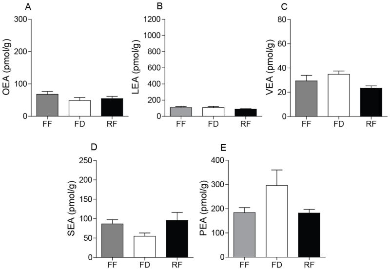
The gastrointestinal tract plays a critical role in the regulation of energy homeostasis by initiating neural and hormonal responses to the ingestion of nutrients. In addition to peptide hormones, such as cholecystokinin (CKK) and peptide YY (PYY), the lipid-derived mediator oleoylethanolamide (OEA) has been implicated in the control of satiety. Previous studies in humans and rodent models have shown that obesity is associated with changes in CCK, PYY and other gut-derived peptide hormones, which may contribute to decreased satiety and increased energy intake. In the present study, we show that small-intestinal OEA production is disrupted in the gut of diet-induced obese (DIO) rats and mice. In lean rodents, feeding or duodenal infusion of Intralipid® or pure oleic acid stimulates jejunal OEA mobilization. This response is strikingly absent in DIO rats and mice. Confirming previous reports, we found that feeding rats or mice a high-fat diet for 7 days is sufficient to suppress jejunal OEA mobilization. Surprisingly, a similar effect is elicited by feeding rats and mice a high-sucrose low-fat diet for 7 days. Collectively, our findings suggest that high fat-induced obesity is accompanied by alterations in the post-digestive machinery responsible for OEA biosynthesis, which may contribute to reduced satiety and hyperphagia.

Keywords: Diet-induced obesity; Dietary fat; Linoleoylethanolamide; Oleoylethanolamide; Satiety.


Figures

Figure 1
Figure 1
Chromatographic separation of OEA and VEA. (A) Representative chromatogram of octadecenoylethanolamides (selected-ion monitoring, SIM, m/z 326). Authentic standards of OEA, VEA, tVEA and EEA were injected (0.5 pmol/injection). (B) Representative SIM chromatogram of octadecenoylethanolamides extracted from rat jejunum mucosa. Five μl of lipid extract prepared from rat jejunum mucosa were injected.
Figure 2
Figure 2
Effects of HFD-induced obesity on FAE mobilization in rat jejunum. Effects of free feeding (FF), 24-h food deprivation (FD), and 30-min refeeding (RF) after 24-h food deprivation on jejunal levels of OEA (A1 and B1), LEA (A2 and B2) and VEA (A3 and B3) SEA (A4 and B4) and PEA (A5 and B5) in lean (A) and DIO (B) rats. Results are expressed as mean ± SEM (n=4–8/feeding condition). ***P<0.001, FF vs. FD and RF vs. FD.
Figure 3
Figure 3
Effects of DIO on levels of octadecenoic acids (oleic and vaccenic) and stearic acid in rat jejunum. Levels of octadecenoic acids (A1 and B1) and stearic acid (A2 and B2) in lean (A) and DIO (B) rats under FF, FD and RF conditions. Oleic and vaccenic acids were not separated under the chromatographic conditions used. Results are expressed as mean ± SEM (n=4–8/feeding condition). *P<0.05, FF vs. FD and RF vs. FD.
Figure 4
Figure 4
Effects on intraduodenal lipid infusion on FAE mobilization in jejunum of DIO rats. Effects of intraduodenal infusion of various macronutrients on jejunal levels of OEA, LEA, VEA and SEA in (A) lean and (B) DIO rats. S, saline; L, Intralipid®; C, sucrose; P, peptone. Rats were deprived of food for 24 h prior to infusions. Infusions were conducted at 0.5 ml/min for 10 min with nutrients equicaloric at 2 kcal/ml. Jejunum mucosa was harvested 30 min after starting the infusion. Results are expressed as mean ± SEM (n=4–5/infusion). **P<0.01 and ***P<0.001, saline vs. Intralipid®.
Figure 5
Figure 5
Effects of intraduodenal oleic acid infusion on FAE mobilization in the jejunum of DIO rats. Intraduodenal infusion of oleic acid (OA) on jejunal levels of OEA, LEA, VEA and SEA in lean (A) and DIO (B) rats. Rats were deprived of food for 24 h prior to infusions. Infusions were conducted at 0.5 ml/min for 10 min with either vehicle (V) or 5% oleic acid emulsion. Jejunum mucosa was harvested 30 min after starting the infusion. Results are expressed as mean ± SEM (n=3–5/infusion). ***P<0.001, vehicle vs. OA.
Figure 6
Figure 6
Feeding-dependent FAE mobilization in the jejunum of rats fed a HFD for 7 days. Levels of OEA (A), LEA (B), VEA (C), SEA (D) and PEA (E) of rats fed a HFD for 7 days. Results are expressed as mean ± SEM (n=5–6/feeding condition).

References

    1. Stanley S, Wynne K, McGowan B, Bloom S. Hormonal regulation of food intake. Physiological reviews. 2005;85:1131–1158. - PubMed
    1. Piomelli D. A fatty gut feeling. Trends in endocrinology and metabolism: TEM. 2013;24:332–341. - PMC - PubMed
    1. Rodriguez de Fonseca F, Navarro M, Gomez R, Escuredo L, Nava F, Fu J, Murillo-Rodriguez E, Giuffrida A, LoVerme J, Gaetani S, Kathuria S, Gall C, Piomelli D. An anorexic lipid mediator regulated by feeding. Nature. 2001;414:209–212. - PubMed
    1. Schwartz GJ, Fu J, Astarita G, Li X, Gaetani S, Campolongo P, Cuomo V, Piomelli D. The lipid messenger OEA links dietary fat intake to satiety. Cell metabolism. 2008;8:281–288. - PMC - PubMed
    1. Astarita G, Rourke BC, Andersen JB, Fu J, Kim JH, Bennett AF, Hicks JW, Piomelli D. Postprandial increase of oleoylethanolamide mobilization in small intestine of the Burmese python (Python molurus) American journal of physiology. Regulatory, integrative and comparative physiology. 2006;290:R1407–1412. - PubMed

Publication types

MeSH terms