

Loss of Function Mutations in NNT Are Associated With Left Ventricular Noncompaction
- PMID: 26025024
- PMCID: PMC4545476
- DOI: 10.1161/CIRCGENETICS.115.001026
Loss of Function Mutations in NNT Are Associated With Left Ventricular Noncompaction
Abstract
Background: Left ventricular noncompaction (LVNC) is an autosomal-dominant, genetically heterogeneous cardiomyopathy with variable severity, which may co-occur with cardiac hypertrophy.
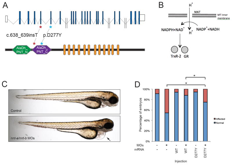
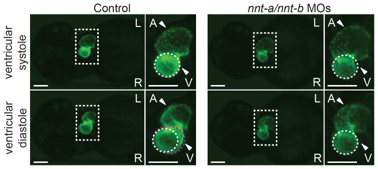
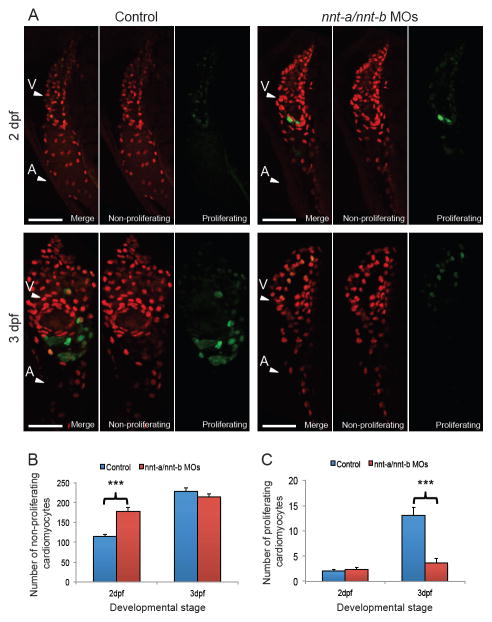
Methods and results: Here, we generated whole exome sequence data from multiple members from 5 families with LVNC. In 4 of 5 families, the candidate causative mutation segregates with disease in known LVNC genes MYH7 and TPM1. Subsequent sequencing of MYH7 in a larger LVNC cohort identified 7 novel likely disease causing variants. In the fifth family, we identified a frameshift mutation in NNT, a nuclear-encoded mitochondrial protein, not implicated previously in human cardiomyopathies. Resequencing of NNT in additional LVNC families identified a second likely pathogenic missense allele. Suppression of nnt in zebrafish caused early ventricular malformation and contractility defects, probably driven by altered cardiomyocyte proliferation. In vivo complementation studies showed that mutant human NNT failed to rescue nnt morpholino-induced heart dysfunction, indicating a probable haploinsufficiency mechanism.
Conclusions: Together, our data expand the genetic spectrum of LVNC and demonstrate how the intersection of whole exome sequence with in vivo functional studies can accelerate the identification of genes that drive human genetic disorders.
Keywords: genetics; genomics; human; mutation; nonisolated left ventricular noncompaction.
© 2015 American Heart Association, Inc.
Conflict of interest statement
Figures
Comment in
-
Malformations of the Left Ventricle: What Comes First: Form or Function?Circ Cardiovasc Genet. 2015 Aug;8(4):537-40. doi: 10.1161/CIRCGENETICS.115.001189. Circ Cardiovasc Genet. 2015. PMID: 26286726 No abstract available.
References
-
- Finsterer J. Cardiogenetics, neurogenetics, and pathogenetics of left ventricular hypertrabeculation/noncompaction. Pediatr Cardiol. 2009;30:659–681. - PubMed
-
- Sarma RJ, Chana A, Elkayam U. Left ventricular noncompaction. Prog Cardiovasc Dis. 2010;52:264–273. - PubMed
-
- Chin TK, Perloff JK, Williams RG, Jue K, Mohrmann R. Isolated noncompaction of left ventricular myocardium. A study of eight cases. Circulation. 1990;82:507–513. - PubMed
-
- Stöllberger C, Blazek G, Wegner C, Winkler-Dworak M, Finsterer J. Neuromuscular and cardiac comorbidity determines survival in 140 patients with left ventricular hypertrabeculation/noncompaction. Int J Cardiol. 2011;150:71–74. - PubMed
-
- Pignatelli RH, McMahon CJ, Dreyer WJ, Denfield SW, Price J, Belmont JW, et al. Clinical characterization of left ventricular noncompaction in children: a relatively common form of cardiomyopathy. Circulation. 2003;108:2672–2678. - PubMed
Publication types
MeSH terms
Substances
Grants and funding
LinkOut - more resources
Full Text Sources
Molecular Biology Databases
Miscellaneous
