

Comparative Analysis of T Cell Imaging with Human Nuclear Reporter Genes
- PMID: 26025962
- PMCID: PMC4511596
- DOI: 10.2967/jnumed.115.159855
Comparative Analysis of T Cell Imaging with Human Nuclear Reporter Genes
Abstract
Monitoring genetically altered T cells is an important component of adoptive T cell therapy in patients, and the ability to visualize their trafficking/targeting, proliferation/expansion, and retention/death using highly sensitive reporter systems that do not induce an immunologic response would provide useful information. Therefore, we focused on human reporter gene systems that have the potential for translation to clinical studies. The objective of the in vivo imaging studies was to determine the minimum number of T cells that could be visualized with the different nuclear reporter systems. We determined the imaging sensitivity (lower limit of T cell detection) of each reporter using appropriate radiolabeled probes for PET or SPECT imaging.
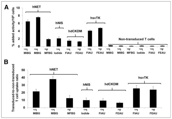
Methods: Human T cells were transduced with retroviral vectors encoding for the human norepinephrine transporter (hNET), human sodium-iodide symporter (hNIS), a human deoxycytidine kinase double mutant (hdCKDM), and herpes simplex virus type 1 thymidine kinase (hsvTK) reporter genes. After viability and growth were assessed, 10(5) to 3 × 10(6) reporter T cells were injected subcutaneously on the shoulder area. The corresponding radiolabeled probe was injected intravenously 30 min later, followed by sequential PET or SPECT imaging. Radioactivity at the T cell injection sites and in the thigh (background) was measured.
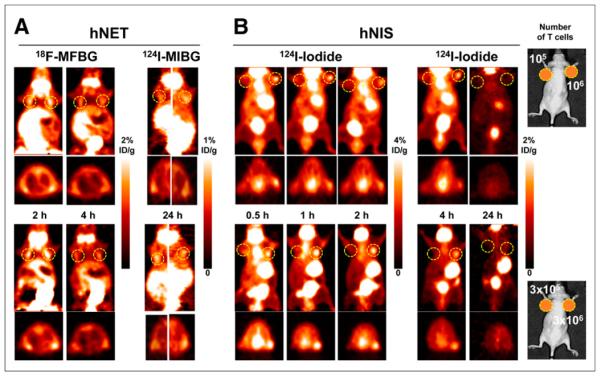
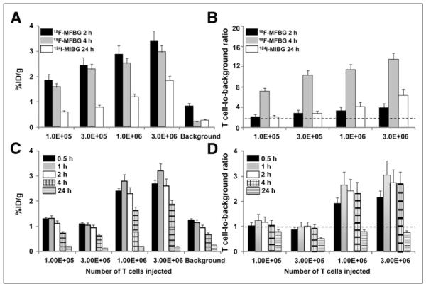
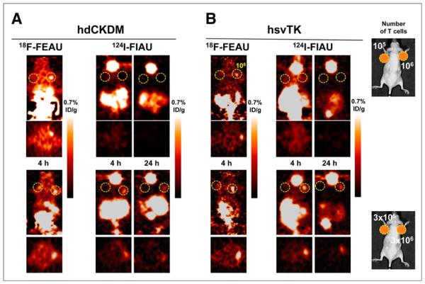
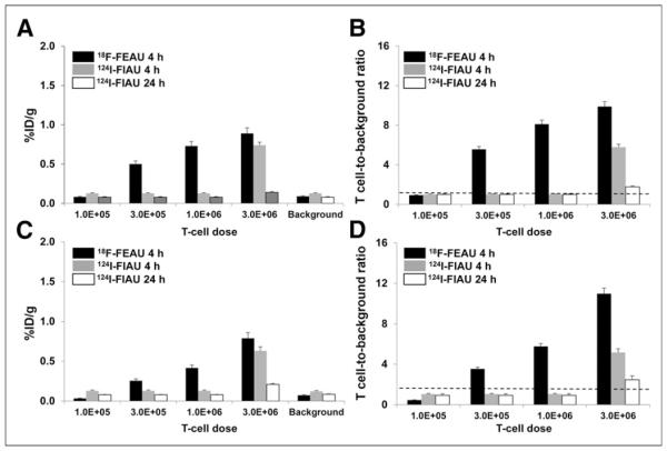
Results: The viability and growth of experimental cells were unaffected by transduction. The hNET/meta-(18)F-fluorobenzylguanidine ((18)F-MFBG) reporter system could detect less than 1 × 10(5) T cells because of its high uptake in the transduced T cells and low background activity. The hNIS/(124)I-iodide reporter system could detect approximately 1 × 10(6) T cells; (124)I-iodide uptake at the T cell injection site was time-dependent and associated with high background. The hdCKDM/2'-(18)F-fluoro-5-ethyl-1-β-d-arabinofuranosyluracil ((18)F-FEAU) and hsvTK/(18)F-FEAU reporter systems detected approximately 3 × 10(5) T cells, respectively. (18)F-FEAU was a more efficient probe (higher uptake, lower background) than (124)I-1-(2-deoxy-2-fluoro-1-d-arabinofuranosyl)-5-iodouracil for both hdCKDM and hsvTK.
Conclusion: A comparison of different reporter gene-reporter probe systems for imaging of T cell number was performed, and the hNET/(18)F-MFBG PET reporter system was found to be the most sensitive and capable of detecting approximately 35-40 × 10(3) T cells at the site of T cell injection in the animal model.
Keywords: PET; T cells; imaging; reporter genes.
© 2015 by the Society of Nuclear Medicine and Molecular Imaging, Inc.
Figures
References
-
- Koehne G, Doubrovin M, Doubrovina E, et al. Serial in vivo imaging of the targeted migration of human HSV-TK-transduced antigen-specific lymphocytes. Nat Biotechnol. 2003;21:405–413. - PubMed
-
- Su H, Chang DS, Gambhir SS, Braun J. Monitoring the antitumor response of naive and memory CD8 T cells in RAG1−/− mice by positron-emission tomography. J Immunol. 2006;176:4459–4467. - PubMed
-
- Choi SR, Zhuang ZP, Chacko AM, et al. SPECT imaging of herpes simplex virus type1 thymidine kinase gene expression by 123I-FIAU(1) Acad Radiol. 2005;12:798–805. - PubMed
Publication types
MeSH terms
Substances
Grants and funding
LinkOut - more resources
Full Text Sources
Other Literature Sources