. 2015 Jul 13;43(12):5687-98.
doi: 10.1093/nar/gkv566. Epub 2015 May 30.

Outwitting EF-Tu and the ribosome: translation with d-amino acids

Affiliations

Outwitting EF-Tu and the ribosome: translation with d-amino acids

John Achenbach et al. Nucleic Acids Res. .

Abstract

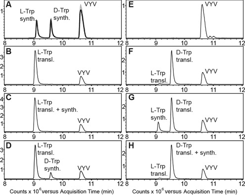
Key components of the translational apparatus, i.e. ribosomes, elongation factor EF-Tu and most aminoacyl-tRNA synthetases, are stereoselective and prevent incorporation of d-amino acids (d-aa) into polypeptides. The rare appearance of d-aa in natural polypeptides arises from post-translational modifications or non-ribosomal synthesis. We introduce an in vitro translation system that enables single incorporation of 17 out of 18 tested d-aa into a polypeptide; incorporation of two or three successive d-aa was also observed in several cases. The system consists of wild-type components and d-aa are introduced via artificially charged, unmodified tRNA(Gly) that was selected according to the rules of 'thermodynamic compensation'. The results reveal an unexpected plasticity of the ribosomal peptidyltransferase center and thus shed new light on the mechanism of chiral discrimination during translation. Furthermore, ribosomal incorporation of d-aa into polypeptides may greatly expand the armamentarium of in vitro translation towards the identification of peptides and proteins with new properties and functions.


Figures

Figure 1.
Figure 1.
l-aa-tRNA and d-aa-tRNA bound to EF-Tu and the ribosomal A-site. (A) Structure of a TC l-Phe-tRNAPhe-EF-Tu-GDPNP. (B) Zoom into the EF-Tu amino acid binding pocket. Hydrogen bonds are indicated as dashed lines. (C, D) Models of d-Phe-tRNAPhe bound to EF-Tu. In (C), the d-Phe side chain faces steric constraints or clashes within the pocket, whereas in (D), clashes are avoided but interactions between EF-Tu and the d-Phe α-amino group are lost. (E) Cytosol side or backside view of the 50S ribosomal subunit. (F) the ribosomal PTC with an l-Phe-tRNA in the A-site. The α-amino group makes several interactions and starts a nucleophilic attack on the peptidyl-tRNA ester carbon for peptide bond formation (red arrow). (G,H) Models of d-Phe-tRNA in the A-site. In (G), the side chain clashes with U2585 and U2506, whereas in (H), the side chain can be accommodated but the α-amino group is turned into an unfavorable position for peptidyl transfer. Coordinates from PDBs 1TTT (41) and 1VQN (45).
Figure 2.
Figure 2.
TC formation with wild-type EF-Tu•GTP and aa-tRNA. The esterified amino acids are ordered in a descending order of binding energy contribution according to (47) (* data for l-His were unavailable, we arbitrarily put it to the left). (A) Upper panel: EMSA with native (n) and unmodified (u) tRNAGly acylated with weakly contributing l-aa. Lower panel: relative quantification of TCs versus unbound tRNA. (B–F) Evaluation of EMSAs with several tRNAs acylated with canonical aa. (B) unmodified tRNAGlyu, (C) transplanted tRNAGlytp, (D) native tRNATyrn, (E) unmodified tRNATyru, (F) transplanted tRNATyrtp. (G, H) Evaluation of EMSAs with tRNAGlyu (G) and tRNATyrtp (H) misacylated with d-aa (see also Supplementary Figure S1).
Figure 3.
Figure 3.
Incorporation of d-aa into peptides. (A) Tricine-SDS-PAGE analyses of single, double and triple incorporation of d-aa. Templates ‘G1’, ‘G2’ and ‘G3’ were translated in the presence of wild-type EF-Tu and tRNAGlyu misacylated with the indicated d- or l-amino acid. (B–D) Relative incorporation efficiencies of d-aa and l-aa from single (B), double (C) and triple (D) incorporation experiments. The intensities of full-length product bands were assessed and background was subtracted globally. The control signal was subtracted from the d-aa signal and the resulting value divided by the l-aa signal. Data are shown as means of at least two independent experiments (n > 2 where indicated), error bars show the range of determined data.
Figure 4.
Figure 4.
The effect of tRNA properties on d-aa incorporation. Single incorporation assays with four different tRNAs misacylated with six different l- and d-amino acids were performed in independent duplicates (triplicates for Lys). Template ‘G1’ was used for translation with tRNAGlyu (high EF-Tu and A-site affinity) and tRNAGlytp (low EF-Tu affinity, high A-site affinity) and template ‘O’ for tRNATyru (low EF-Tu and A-site affinity) and tRNATyrtp (high EF-Tu affinity, low A-site affinity). The anticodon on both tRNATyr was changed to UCA to enable opal stop codon suppression. (A) Autoradiograms of the gels. Remarkable differences in the d-aa incorporation efficiencies depending on EF-Tu affinity become apparent. (B) Signal intensities normalized to the respective reaction with l-aa-tRNAGlyu (lane 2 in (A)) are plotted. Control signals were subtracted from d-aa reaction signals prior to normalization. For clarity, the y-axis scaling is compressed above 150%. Error bars show the range of determined data.
Figure 5.
Figure 5.
Tricine-SDS-PAGE analyses of single D-aa incorporation with wild-type EF-Tu and EF-Tu N273S. Template ‘G1’ was translated in the presence of tRNAGlyu misacylated with selected d- and l-amino acids. (A) Autoradiograms of the gels. (B) Quantification of full-length product bands relative to bands from l-reactions with wild-type EF-Tu (lane 2 in (A)). EF-Tu N273S is inferior to the wild-type in all cases.
Figure 6.
Figure 6.
LC-MS analysis of translation products. The graphs show merged extracted ion chromatograms of the most intense charge states of the full-length peptides [(M+5H)5+: 539.6 m/z, (M+4H)4+: 674.3 m/z, (M+3H)3+: 898.7m/z; asymmetric peak detection (−0.3 Da/+0.7 Da); calculated monoisotopic mass (2693.3 Da) and of the synthetic tripeptide VYV (380.2 m/z). (A) Overlay of three standards each run before (grey) and after the samples (50 ng per peptide) (black). (B) Translated l-Trp peptide (∼350 ng) with VYV. (C) Translated l-Trp peptide with synthetic l-Trp peptide and VYV. The l-Trp peptides co-elute. (D) Translated l-Trp peptide with synthetic d-Trp peptide and VYV. The peptides are separated. (E) Negative control with VYV. (F) Translated d-Trp peptide (∼240 ng) with VYV. The d-Trp peptide elutes at the expected retention time, a minor peak corresponding to the l-Trp peptide (∼7 ng) is also detected. (G) Translated d-Trp peptide with synthetic l-Trp peptide and VYV. The peptides are separated. (H) Translated d-Trp peptide with synthetic d-Trp peptide and VYV. The d-Trp peptides co-elute.

References

    1. Kreil G. D-amino acids in animal peptides. Annu. Rev. Biochem. 1997;66:337–345. - PubMed
    1. Luo L., Kohli R.M., Onishi M., Linne U., Marahiel M.A., Walsh C.T. Timing of epimerization and condensation reactions in nonribosomal peptide assembly lines: kinetic analysis of phenylalanine activating elongation modules of tyrocidine synthetase B. Biochemistry. 2002;41:9184–9196. - PubMed
    1. Erspamer V., Melchiorri P., Falconieri-Erspamer G., Negri L., Corsi R., Severini C., Barra D., Simmaco M., Kreil G. Deltorphins: a family of naturally occurring peptides with high affinity and selectivity for delta opioid binding sites. Proc. Natl Acad. Sci. U.S.A. 1989;86:5188–5192. - PMC - PubMed
    1. Chen S., Gfeller D., Buth S.A., Michielin O., Leiman P.G., Heinis C. Improving binding affinity and stability of peptide ligands by substituting glycines with D-amino acids. Chembiochem. 2013;14:1316–1322. - PubMed
    1. Soutourina J., Plateau P., Blanquet S. Metabolism of D-aminoacyl-tRNAs in Escherichia coli and Saccharomyces cerevisiae cells. J. Biol. Chem. 2000;275:32535–32542. - PubMed

Publication types

MeSH terms